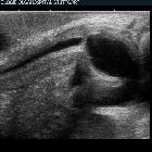
posterior urethral valve


 nicht verwechseln mit: Ureterklappen
Posterior urethral valves (PUVs), also referred as congenital obstructing posterior urethral membranes (COPUM), are the most common congenital obstructive lesion of the urethra and a common cause of obstructive uropathy in infancy.
Epidemiology
Posterior urethral valves are congenital and only seen in male infants . The estimated incidence is at ~1 in 10,000-25,000 live births with a higher rate of occurrence in utero.
Clinical presentation
Clinical presentation depends on the severity of obstruction. In severe obstruction, the diagnosis is usually made antenatally. The fetus will be small for gestational age and ultrasound examination will demonstrate oligohydramnios and associated abnormalities (see below) . In less severe cases, the diagnosis is often not apparent until early infancy. Urinary tract infections are common in this group .
Pathology
Posterior urethral valves result from the formation of a thick, valve-like membrane from a tissue of Wolffian duct origin (failure of regression of the mesonephric duct ) that courses obliquely from the verumontanum to the most distal portion of the prostatic urethra. This is thought to occur in early gestation (5-7 weeks ). The valve is a diaphragm with a central pinhole, however as it is more rigid along its line of fusion it gradually distends and becomes distended into a bilobed sail-like or windsock-like structure .
According to Young's classification, there are three types of posterior urethral valves :
- type 1
- most common
- occurs when the two mucosal folds extend anteroinferiorly from bottom of verumontanum and fuse anteriorly at lower level
- type 2
- rare
- no longer considered as a valve but a normal variant
- mucosal folds extend along posterolateral urethral wall from ureteric orifice to verumontanum
- type 3
- circular diaphragm with central opening in membranous urethra
- located below the verumontanum and occurs due to abnormal canalization of urogenital membrane
- sometimes referred to as Cobb's collar
Genetics
The vast majority of cases are sporadic, although rare examples of posterior urethral valves occurring in families have been reported .
Associations
Posterior urethral valves are also seen in association with other congenital abnormalities including :
- chromosomal abnormalities, e.g. Down syndrome
- bowel atresia
- craniospinal defects
Radiographic features
Ultrasound
Antenatal ultrasound
On antenatal ultrasound, the appearance is that of:
- marked distention and hypertrophy of the bladder
- hydronephrosis and hydroureter may or may not be present
- in severe cases oligohydramnios and renal dysplasia (assessing the degree of renal dysplasia is difficult antenatally, although some authors believe that significantly increased echogenicity of the kidneys is an indication of poor function )
- keyhole sign may be seen on ultrasound due to the distention of both the bladder and the urethra immediately proximal to the valve
Unfortunately, such findings are generally not seen before 26 weeks of gestation, and as such are not frequently identified on routine morphology screening, usually carried out around 18 weeks gestation .
Postnatal ultrasound
Following birth, findings are the same as those on antenatal ultrasound. Patients who present after birth usually have less severe obstruction, and so the features may be less evident:
- bladder is typically thick-walled and trabeculated with an elongated and dilated posterior urethra (keyhole sign)
- hydronephrosis (most commonly), although it is important to note this is absent in up to 10% of cases
- kidneys may be hyperechoic with loss of normal corticomedullary differentiation, a manifestation of renal dysplasia
- examination of posterior urethra can be performed longitudinally through the perineum
- ideally performed during micturition (which may take some patience) at which time the proximal urethra can be seen to dilate
- diameter >6 mm considered abnormal, and is highly specific and sensitive (sensitivity 100%, specificity 89%, positive predictive value 88%)
- the abnormal valve may be seen as an echogenic line
In some cases high pressure generated during attempted micturition can result in rupture of the collecting system with accumulations of urine in various compartments, including :
- rupture of a calyceal fornix: result in pararenal urinomas, seen as anechoic fluid collections around the kidney
- intraperitoneal bladder rupture: intraperitoneal fluid, indistinguishable ultrasonographically from ascites
Voiding cystourethrogram (VCUG)
Voiding cystourethrogram (VCUG) is the best imaging technique for the diagnosis of posterior urethral valves. The diagnosis is best made during the micturition phase in lateral or oblique views, such that the posterior urethra can be imaged adequately . Findings include :
- dilatation and elongation of the posterior urethra (the equivalent of the ultrasonographic keyhole sign)
- linear radiolucent band corresponding to the valve (only occasionally seen)
- vesicoureteral reflux (VUR): seen in 50% of patients
- bladder trabeculation / diverticula
Treatment and prognosis
Antenatal treatment is possible, consisting of vesicoamniotic shunting (allowing urine to exit the bladder via the shunt, bypassing the obstructed urethra). Essentially this procedure consists of a supra-pubic catheter performed under ultrasound guidance. The efficacy of this procedure is controversial, as often despite this significant renal and pulmonary morbidity exist .
Postnatally, definitive treatment is simple and involves transurethral ablation of the offending valve .
The overall prognosis is most affected by the degree and duration of obstruction. Severe cases with obstructive cystic renal dysplasia, oligohydramnios and pulmonary hypoplasia are often incompatible with life .
History and etymology
The initial three level classification system of posterior urethral valves was proposed in 1919 by Hugh Hampton Young (1870–1945). Young also developed a transurethral punch instrument to treat these valves .
Differential diagnosis
In the correct age group and with clear dilatation of the posterior urethra there is usually little differential other than urethral atresia, which is far less common .
When only the bladder is clearly abnormal thick-walled and trabeculated, other conditions to be considered include :
Siehe auch:
und weiter:
- Eagle-Barrett syndrome
- genitourinary curriculum
- Harnblasendivertikel
- Oligohydramnion
- fetal hydronephrosis
- fetal pyelectasis
- Vesikoureteraler Reflux
- Potter-Sequenz
- Obstruktive Uropathie
- Anhydramnion
- Blasenentleerungsstörung
- megacystis microcolon intestinal hypoperistalsis syndrome
- Harnröhrenstriktur
- causes of urinary bladder diverticulae
- urethral agenesis
- kongenitale Anomalien der Urethra
- fetale Megazystis
- kongenitaler Megaureter
- Schlüssellochzeichen
- Schlüssellochzeichen posteriore Urethralklappe
- kongenitale Urethrastenose

 Assoziationen und Differentialdiagnosen zu Urethralklappe:


