second branchial cleft cyst


Second branchial cleft cysts are a cystic dilatation of the remnant of the second branchial cleft (see branchial apparatus), and along with second branchial fistulae and sinuses accounts for 95% of all branchial cleft anomalies.
Clinical presentation
Although a congenital abnormality, they tend to present in early adulthood (10-40 years of age) often after minor trauma or infection. Second branchial cleft sinus or fistulas, on the other hand, present earlier. Fistulas extend from the skin surface anterior to the middle of the sternocleidomastoid muscle, pass between the internal and external carotid arteries and eventually drain into the tonsillar fossa.
Typically, second branchial cleft cysts present as a rounded swelling just below the angle of mandible, anterior to the sternocleidomastoid (although the position is variable - see classification below).
Pathology
The cyst is typically filled with mucoid material, is well circumscribed and other than presenting as localized swelling, is asymptomatic. However, if infected, surrounding fat stranding and skin changes are evident.
Location
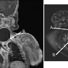
Cysts can occur anywhere along the course of the second branchial apparatus, from the pharyngeal wall to the skin, as it passes laterally and inferiorly between the internal and external carotid arteries. The angle of the mandible is a common location.
Classification
Radiographic features
Ultrasound
- sharply demarcated
- posterior acoustic enhancement: 70%
- imperceptible walls: 82%
- echogenicity is variable
- anechoic: 41%
- homogeneously hypoechoic with internal debris: 24%
- pseudo solid: 12%
- heterogeneous: 23%
CT
- rounded or spheric, sharply circumscribed
- fluid density centrally
- thin wall
- extension of the cyst wall between the internal and external carotid arteries just above the carotid bifurcation (features sometimes referred to as the notch sign, tail sign or beak sign ), is highly suggestive of the diagnosis but not pathognomonic
MRI
- T1: variable signal dependent on protein content
- high protein content: high signal
- low protein content: low signal
- T2: usually high signal
- T1 C+ (Gd): no enhancement in uncomplicated lesions
Treatment and prognosis
Complications
- superimposed infection
Differential diagnosis
- paramedian thyroglossal duct cyst
- cystic lymph nodes
- necrotic nodal metastases, especially squamous cell carcinoma and papillary thyroid cancer
- tuberculous adenitis
- vascular lesions
- jugular vein thrombosis
- mycotic aneurysm of the neck
- neurogenic tumors
- cervical dermoid cyst
- cavernous lymphangioma
- thyroid nodule/cyst
Siehe auch:
- Schwannom
- zystische Lymphknoten
- Neurofibrom
- Ductus thyreoglossus Zyste
- lymphoepitheliale Zyste der Glandula parotis
- zervikale bronchogene Zyste
- benignes Ganglioneurom
- tuberkulöse Halslymphknoten
- Halsfistel
- Kiemenbogenzyste
- Zyste oder Fistel des vierten Kiemenbogens
- Kiemenbogen
und weiter:
- laterale Halszyste
- fatty nodal metaplasia
- differential diagnosis of paediatric cervical lesions
- Zyste oder Fistel des dritten Kiemenbogens
- cystic cervical mass adjacent to the angle of mandible
- Mundbodenzyste (Ranula)
- notch sign
- second branchial cleft cyst classification
- Fistel des zweiten Kiemenbogens
- infizierte Zyste oder Fistel des zweiten Kiemenbogens


 Assoziationen und Differentialdiagnosen zu Zyste oder Fistel des zweiten Kiemenbogens: