Susac syndrome


Susac syndrome (SS), also known as SICRET syndrome (small infarctions of cochlear, retinal and encephalic tissue), is a rare syndrome typically affecting young to middle-age women that is clinically characterized by the triad of acute or subacute encephalopathy, bilateral sensorineural hearing loss, and branch retinal arterial occlusions.
Epidemiology
The condition more commonly affects females around the 2 to 4 decades . Approximately three females are affected for every male case .
Clinical presentation
It consists of a clinical triad of:
- acute or subacute encephalopathy which could manifest in a broad spectrum of symptoms, e.g. memory impairment, confusion, behavioral disturbances, ataxia, dysarthria, paranoid psychosis, and headaches
- sensorineural hearing loss at low and medium frequencies
- branch retinal artery occlusions leading to scotomata and vision distortion
All clinical features may not be present at the same time, and may, in fact, fluctuate over a 1-2 year period, which often leads to a delay in diagnosis .
Branch retinal artery occlusions (BRAO) should be sought by fluorescein angiogram as it may help to differentiate from multiple sclerosis .
Diagnostic criteria
Diagnostic criteria have been proposed in 2016 that divide patients into definite and probable diagnosis of Susac syndrome based on the presence of certain clinical and imaging criteria . The criteria are:
- i. symptoms and clinical findings:
- new cognitive impairment
- behavioral changes
- new focal neurological symptoms
- headache (within 6 months of diagnosis; migrainous or oppressive in character)
- ii. imaging features
- rounded T2 hyperintense "snowball lesions"
- at least one in the corpus callosum
- leptomeningeal enhancement
- well demarcated grey matter enhancing on T1 weighted scans
- i. no clinical findings or symptoms required
- ii. ophthalmological examinations
- branch retinal artery occlusions (BRAO)
- arterial wall hyperfluorescence (AWH)
- sectorial damage is seen on optical coherence tomography (OCT)
- i. symptoms and clinical findings:
- new tinnitus
- new hearing loss
- new peripheral vertigo
- ii. examination of inner ear function
- audiogram supporting hearing loss
- vertigo supported by caloric testing and vestibular evoked myogenic responses
A definite diagnosis of Susac syndrome requires fulfillment of all three criteria and their subcriteria, in other words, 1-i, 1-ii, 2-ii, 3-i and 3-ii.
A probable diagnosis of Susac syndrome can be made if 2 out of the three criteria are met.
Pathology
The primary pathology is likely to be an autoimmune endothelialopathy (microangiopathy) affecting pre-capillary arterioles, with subsequent embolization, of the brain, retina, and inner ear . Hence, microinfarcts associated with endothelial proliferation and hypertrophy, basement membrane thickening, and thickening of precapillary arteriolar adventitia with loss of internal elastic lamina . Demyelination is not a pathological hallmark of Susac syndrome, in contrast with multiple sclerosis .
Markers
Anti-endothelial cell antibodies (AECAs) have been reported as being a promising marker in some studies, but these are not yet considered to be specific to the syndrome .
Radiographic features
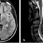
MRI
Characteristic radiographic features are present even if all components of the clinical triad have not yet manifested . There tend to be multiple, small white matter lesions which have a predilection for the corpus callosum .
Callosal lesions are considered almost pathognomonic and have many important characteristic features:
- typically involve the central fibers of the callosal body and splenium without abuting the callosal undersurface (with relative sparing of the periphery)
- lesions are small (3-7 mm) and resembling :
- 'snowballs' on T2 and FLAIR when acute
- 'punched out' holes on T1 when chronic
- lesions, best appreciated on sagittal views, depending on the sequence used
- the roof of the corpus callosum is also frequently involved, rather than the callososeptal interface (which is more typical of multiple sclerosis), resulting in 'icicle' or linear 'spoke' lesions that look to hang from the roof of the corpus callosum
In addition to the corpus callosum, lesions can also involve the periventricular white matter, centrum semiovale, cerebellum, brainstem, and middle cerebellar peduncles . A 'string of pearls' appearance due to punctate microinfarcts involving the internal capsule has been described .
Signal characteristics of all of these lesions include:
- T1: lesions are low signal, especially in the chronic stage (see T1 black holes)
- T1 C+ (Gd): lesions frequently enhance during the acute stage
- T2/FLAIR: lesions are high signal
- DWI/ADC: normal, although T2 shine-through may be appreciated on DWI
Treatment and prognosis
Although Susac syndrome generally has a monophasic and self-limiting course, case series-level evidence suggests management of the acute episode with immunosppressants such as prednisolone . Intratympanic injection of dexamethasone has also shown to help with hearing loss, although in severe cases cochlear implants should be considered . Some patients may have residual neurological sequelae, especially related to retinal infarction .
History and etymology
Initially described by John O. Susac (1940-2012), an American neurologist and neuro-ophthalmologist, and his colleagues in their 1979 seminal paper describing two patients with the classic clinical triad . The eponym was famously first used in 1986, when a patient was presented in a neuro-ophthalmology conference with the same triad that Susac et al. had described, prompting one member of the conference to exclaim “this is Susac’s syndrome” .
Differential diagnosis
Considerations on the basis of pure radiographic (MRI) appearances include:
- demyelinating conditions
- multiple infarcts caused by
- thromboembolic disease
- CADASIL
- vasculitides
- systemic lupus erythematosus (SLE) related vasculitis
- transient lesion of splenium of corpus callosum: shows restricted diffusion and no enhancement.
- marchiafava-bignami disease
Siehe auch:
- Vaskulitis
- Encephalomyelitis disseminata
- systemischer Lupus Erythematodes
- CADASIL
- Akute disseminierte Enzephalomyelitis
- Balkendefekte
und weiter:


 Assoziationen und Differentialdiagnosen zu Susac syndrome: