Thymic tumors

Role of
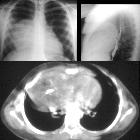
different imaging modalities in the evaluation of normal and diseased thymus. a Chest X-ray PA view shows a near-total opacification of the left hemithorax and right upper lung zone merging with the mediastinum. b Axial CT chest mediastinal window shows a large fat attenuating mass with thin hypodense fibrous septae. c MRI lung T1 WI shows the hyper-intense signal of the mass confirming its fatty nature. Imaging diagnosis and histopathologic assessment revealed “thymolipoma”

Mikronoduläres
Thymom mit lymphoidem Stroma. Hier Computertomographie axial.

Malignes
Thymom / Thymuskarzinom in der Computertomographie (axial). Die Histologie erbrachte ein primäres Karzinom des Thymus vom Plattenepithelial-Typ, dem häufigsten Typ des Thymuskarzinoms. Das CT zeigt eine große, nicht scharf begrenzte Formation im vorderem Mediastinum mit Verdrängung der Lunge und Zeichen einer möglichen Infiltration in Nachbarstrukturen. Inhomogene Kontrastierung. Zysten oder Verkalkungen fanden sich in diesem Fall nicht.

Malignes
Thymom / Thymuskarzinom in der Computertomographie (coronar). Die Histologie erbrachte ein primäres Karzinom des Thymus vom Plattenepithelial-Typ, dem häufigsten Typ des Thymuskarzinoms. Das CT zeigt eine große, nicht scharf begrenzte Formation im vorderem Mediastinum mit Verdrängung der Lunge und Zeichen einer möglichen Infiltration in Nachbarstrukturen. Inhomogene Kontrastierung. Zysten oder Verkalkungen fanden sich in diesem Fall nicht.

Thymuskarzinom
in der Computertomographie mit Kontrastmittel. Der maligne Charakter wird durch die ummauernde Ausdehnung und unscharfe (infiltrierende!?) Begrenzung deutlich. 1 : Das Karzinom 2 : V. cava superior 3 : Truncus brachiocephalicus 4 : Linke A. subclavia und A. carotis communis 5 : Aortenbogen 6 : Sternum

Cavernous
hemangioma in the thymus: a case report. Enhanced chest computed tomography (CT) scan showing a anterior mediastinal tumor in the left lobe of thymus, 2.0 × 1.2 × 1.8 cm in size, and b vein running in the thymus in communication with the tumor (arrow)

Primary
neoplasms of the thymus • Thymoma - invasive - Ganzer Fall bei Radiopaedia

Primary
neoplasms of the thymus • Thymoma - Ganzer Fall bei Radiopaedia

Primary
neoplasms of the thymus • Thymolipoma - Ganzer Fall bei Radiopaedia

Primary
neoplasms of the thymus • Thymoma - Ganzer Fall bei Radiopaedia

Role of
different imaging modalities in the evaluation of normal and diseased thymus. Sixty-year-old male patient complaining of dyspnea. a Reconstruction image showing right-sided opacity not silhouetting the cardiac border. b–d Contrast-enhanced axial CT chest mediastinal and lung windows showing anterior mediastinal fluid attenuating non-enhancing cystic lesion. It shows a thin rim with no enhancement. CT diagnosis and histopathological confirmation revealed “thymic cyst”
Although primary tumors of the thymus are rare, they are the most common causes of a neoplasm of the anterosuperior mediastinum .
- thymoma (staging)
- one-third are benign
- two-thirds are malignant
- invasive thymoma (most)
- thymic carcinoma (rare)
- thymolipoma/thymoliposarcoma
- thymic cyst
- congenital (contains thymic tissue in wall)
- secondary to thoracotomy
- following chemotherapy or radiotherapy for mediastinal tumors
- inflammatory
- benign thymic hyperplasia
- thymic carcinoid
- primary thymic lymphoma
Siehe auch:
- Thymus
- Thymom
- Thymuskarzinom
- Tumoren des vorderen oberen Mediastinums
- Thymuslipom
- Thymushyperplasie
- invasives Thymom
- Thymuszyste
- thymic epithelial tumours
- neuroendokriner Tumor des Thymus
- Teratom des Thymus
- Metastasen im Thymus
- Karzinoid des Thymus
- primäres Lymphom des Thymus
und weiter:

 Assoziationen und Differentialdiagnosen zu Tumoren des Thymus:


