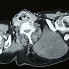
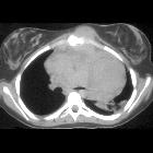
thymolipoma


Thymolipoma is a rare, benign anterior mediastinal mass of thymic origin, containing both thymic and mature adipose tissue.
Epidemiology
Thymolipomas comprise ~5% (range 2-9%) of all thymic neoplasms, but are less common than a mediastinal lipoma of non-thymic origin. There is no recognized sex predilection and reported age range between 3-56 years, with a mean age of 22 years.
Clinical presentation
Most thymolipomas are asymptomatic and found incidentally, often due to imaging of respiratory tract infection. Symptoms, when present are attributable to displacement of mediastinal structures. Approximately 25% complain of non-specific symptoms such as cough, dyspnea and chest pain, which may or may not be the result of the mass.
Pathology
Thymolipomas are composed of a mixture mature adipose tissue with islands of thiymic tissue. The etiology remains uncertain, with hypotheses proposed including
- true neoplasm of the thymus
- variant of a thymoma
- hyperplasia of mediastinal fat
- neoplasm of mediastinal fat which engulfs thymic tissue
Irrespective of cause, these tumors are slow-growing, only gradually increasing in size and are usually large at the time of diagnosis.
Associations
Unlike thymomas, thymolipomas are only occasionally associated with myasthenia gravis . In addition, they are also described in association with
Location
They occur mostly in the cardiophrenic angles.
Radiographic features
A thymolipoma can grow to a very large size before discovery.
Plain radiograph
Typically these tumors appear as large anterior mediastinal masses. The larger tumors tend to hang down one or either side of the pericardium, and being soft, they mold themselves to the adjacent mediastinum and diaphragm and often mimic cardiomegaly . The predominantly fat density can be difficult to identify on plain radiography, however in larger masses that abut the diaphragm, the diaphragm can still be seen.
CT
On CT thymolipomas typically appears almost entirely fatty with some areas of inhomogeneous soft tissue density that represent thymic tissue.
MRI
Thymolipomas have fat and soft tissue signal characteristics:
- T1: typically the adipose tissue within the tumor returns high T1 signal and thymic component a more intermediate T1 signal
- STIR FS: shows complete suppression; consistent with subcutaneous fat
Treatment and prognosis
Surgical local resection is curative; there are no reports of recurrence, metastasis or mortality.
History and etymology
First reported by Lange in 1916. The term was coined by Hall in 1948.
Differential diagnosis
The differential on plain radiography is that of an anterior mediastinal mass. In larger tumors which 'hang' down, causes of cardiomegaly should also be considered.
On CT the differential is much narrower and includes
- mediastinal lipomatosis
- liposarcoma
- overgrowth of cardiophrenic fat pad
- in some situations certain form of focal thymic hyperplasia may have some imaging overlap
- thymoliposarcoma
Siehe auch:
- Thymus
- Teratom
- Liposarkom
- Lymphom
- Morbus Basedow
- Thymom
- Tumoren des vorderen oberen Mediastinums
- Hamartom
- Kardiomegalie
- Morbus Hodgkin
- fetthaltige mediastinale Raumforderungen
- Aplastische Anämie
- Tumoren des Thymus
- Thymusrest
und weiter:

 Assoziationen und Differentialdiagnosen zu Thymuslipom:


