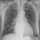
unilateral hyperlucent hemithorax (mnemonic)

Röntgenbild
der Lunge bei einem 3-jährigen Kind nach Aspiration einer Erdnuss: Die linke Lunge ist durch einen Ventilmechanismus durch die Erdnuss deutlich überbläht. Klar erkennbarer Shift des Mediastinums nach rechts (links im Bild).

Fremdkörperaspiration
(Erdnuss oder Apfelstück?) mit Ventilmechanismus und Überblähung der rechten Lunge. Deutlicher Mediastinalshift nach links.

Teenager with
a small deformed right chest wall and a small right hand. CXR PA and lateral shows thinning of the soft tissues over the right chest. The upper right ribs are deformed and there is mild scoliosis. Axial CT without contrast of the chest shows the asymmetry of the thoracic cavity and the concavity of the right chest wall. The right breast is absent, the right pectoralis major and right pectoralis minor muscles are absent, and the right serratus anterior and latissimus dorsi muscles are decreased in size.The diagnosis was Poland syndrome.


Unilateral
hypertranslucent hemithorax • Spontaneous pneumothorax - Ganzer Fall bei Radiopaedia

Respiratory
distress syndrome • Neonatal respiratory distress syndrome, pneumothorax, and PICC line malposition - Ganzer Fall bei Radiopaedia

Unilateral
hypertranslucent hemithorax • Swyer-James syndrome - Ganzer Fall bei Radiopaedia

Unilateral
hypertranslucent hemithorax • Re-expansion pulmonary edema - Ganzer Fall bei Radiopaedia

Gunshot
injuries • Heimlich valve and hemopneumothorax - Ganzer Fall bei Radiopaedia

Poland
syndrome • Poland syndrome - Ganzer Fall bei Radiopaedia

Unilateral
hypertranslucent hemithorax • Mastectomy - Ganzer Fall bei Radiopaedia

Unilateral
hypertranslucent hemithorax • Apical post-radiotheraphy change - Ganzer Fall bei Radiopaedia

Ablatio
mammae rechts mit entsprechend fehlendem Mammaschatten und Hypertransparenz der rechten Thoraxhälfte.

Beyond
bronchitis: a review of the congenital and acquired abnormalities of the bronchus. Obstruction due to foreign body. Frontal (a) and right lateral decubitus (b) images demonstrating mild hyperlucency of the right lung on frontal imaging and lack of volume loss on decubitus imaging, consistent with foreign body obstruction on the right


Imaging of
congenital lung diseases presenting in the adulthood: a pictorial review. Radiological features of congenital lobar hyperinflation in a 42-year-old male presenting with frequent respiratory infections. Chest X-ray shows hyperlucency of the left lung (a). Axial CT image (b) and quantitative 3D reconstruction image (c) demonstrate lobar hyperinflation (c, blue area) and shift to the contralateral side

Unterlappenatelektase
links nach Aspiration eines Pinienkerns. Der linke Oberlappen ist kompensatorisch überbläht.
Mnemonics for a unilateral hyperlucent hemithorax include:
- CRAWLS
- SAFE POEM
- ACROSSS
Mnemonics
CRAWLS
- C: contralateral lung increased density, e.g. supine pleural effusion
- R: rotation
- A: air, e.g. pneumothorax
- W: wall, e.g. chest wall mass, mastectomy, polio, Poland syndrome, surgical removal of the pectoralis major muscle
- L: lungs, e.g. airway obstruction, emphysema, Swyer-James syndrome, unilateral large bullae, large pulmonary embolus
- S: scoliosis
SAFE POEM
- S: Swyer-James syndrome
- A: agenesis (pulmonary)
- F: fibrosis (mediastinal)
- E: effusion (pleural effusion on the contralateral side)
- P: pneumonectomy/pneumothorax
- O: obstruction
- E: embolus (pulmonary)
- M: mucous plug
ACROSSS
- A: air- pneumothorax
artery - pulmonary aplasia or hypoplasia
- C: chest wall - mastectomy, polio, Poland syndrome
- R: rotated film
- O: obstructive causes - airway obstruction, foreign body, unilateral emphysema, or large embolus
- S: scoliosis
- S: surrounding - increased density in contralateral lung, e.g. pleural effusion in the opposite lung in a recumbent patient
- S: Swyer-James syndrome (Macleod syndrome).
See also
Siehe auch:
- Pneumothorax
- Pleuraerguss
- Lungenarterienembolie
- Lungenemphysem
- Westermark-Zeichen
- Bronchiolitis obliterans
- Skoliose
- Herzfehler
- Poland-Syndrom
- kongenitales lobäres Emphysem
- fehlender Mammaschatten in der Thoraxaufnahme nach Mastektomie
- Bronchoventilmechanismus
- fibrosierende Mediastinitis
- bullöses Emphysem
- Swyer-James-Syndrom
- Emphysem
- idiopathisches Lungenemphysem mit riesigen Bullae
- pulmonale Bullae
- agenesis (pulmonary)
- fibrosis (mediastinal)
- Blalock Taussig
und weiter:

 Assoziationen und Differentialdiagnosen zu einseitig vermehrte Transparenz Thorax:
idiopathisches
Lungenemphysem mit riesigen Bullae


