Variant of the aortic arch


Variant anatomy of the aortic arch occurs when there is failure of normal aortic development. It results in a number of heterogenous anomalies of the aorta and its branch vessels.
Gross anatomy
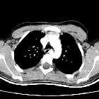
Normally, the aorta ascends in the superior mediastinum to the level of the sternal notch before arching posteriorly and descending in the left hemithorax. The arch gives off three branch vessels, the brachiocephalic (also called the innominate), left common carotid and left subclavian arteries.
Aortic development is a complex process that takes place during the third week of gestation. During development, the two dorsal aortae fuse to form the descending aorta, the ventral aortic limbs fuse to form the aortic sac, the left 4th arch vessel becomes the aortic arch and the right 4th arch vessel becomes atretic distally.
Variant anatomy
Common arch anomalies
When there is departure from normal development, variant anatomy occurs. Commonly, failure of normal regression of the 4th arch vessels results in a double aortic arch or right-sided aortic arch.
Other arch anomalies
- hypoplastic ascending aorta
- coarctation of the aorta
- interrupted aortic arch
- patent ductus arteriosus
- cervical aortic arch
- ductus diverticulum
- circumflex aorta
Branch vessel anomalies
Abnormal formation of the 1st, 2nd and 3rd arch vessels results in abnormal branch vessels:
- bovine arch (commonest, occurring in 10-20% of the population)
- thyroidea ima artery (between 4-10 % of cases)
- variant origin of vertebral arteries (2.5-6% of cases)
- aberrant right subclavian artery (in 0.6% of cases)
- aberrant left subclavian artery (right-sided arch)
- variant aortic branch vessels
- bronchial arteries (case)
Siehe auch:
- Truncus bicaroticus
- Arteria lusoria
- Aortenisthmusstenose
- Aorta
- Arteria carotis communis
- Persistierender Ductus arteriosus
- Abgang der linken Arteria vertebralis aus der Aorta
- rechts descendierende Aorta
- doppelter Aortenbogen
- Normvarianten Truncus brachiocephalicus
- Arteria thyroidea ima
- Varianten Arteria carotis
- vorderes Mediastinum
- vaskuläre Ringe
- zirkumflexer retroösophagealer rechter Aortenbogen
- Kiemenbogenarterien
- fehlende Anlage der Arteria carotis communis beidseits
- Atresie der Aorta
- right arch - left heart
und weiter:


 Assoziationen und Differentialdiagnosen zu Anomalien des Aortenbogens:


