AVM


Arteriovenous
malformation • Ateriovenous malformation of the hand - Ganzer Fall bei Radiopaedia

Arteriovenous
malformation • Intramuscular arteriovenous malformation - Ganzer Fall bei Radiopaedia

Arteriovenous
malformation • Giant scalp arteriovenous malformation - Ganzer Fall bei Radiopaedia

Arteriovenous
malformation • Popliteal arteriovenous malformation - Ganzer Fall bei Radiopaedia

Arteriovenous
malformation • Thenar eminence arteriovenous malformation - Ganzer Fall bei Radiopaedia

Brain
arteriovenous malformation • Cerebral arteriovenous malformation - Ganzer Fall bei Radiopaedia
Arteriovenous
malformation • Congenital arteriovenous malformation of the hand - Ganzer Fall bei Radiopaedia

Arteriovenous
malformation • Arteriovenous malformation - thigh - Ganzer Fall bei Radiopaedia
Arteriovenous
malformation • High-flow arteriovenous malformation of the shoulder - Ganzer Fall bei Radiopaedia

Arteriovenous
malformation • Renal arteriovenous malformation - Ganzer Fall bei Radiopaedia

Arteriovenous
malformation • Arteriovenous malformation of the gluteus - Ganzer Fall bei Radiopaedia

Arteriovenous
malformation • Arteriovenous malformation - face - Ganzer Fall bei Radiopaedia

Arteriovenöse
Malformation des Gehirns in der Computertomographie nativ, also ohne Kontrastmittel. Die erweiterten Gefäßkonvolute sind dennoch hyperdens also hell zu erkennen. Vergleiche auch die MRT-Bilder dieses Falls.

Brain
arteriovenous malformation • Arteriovenous malformation - cerebral - Ganzer Fall bei Radiopaedia

Arteriovenous
malformation with bone lesions. Axial planes figures 6 (T1), 7 (T2FS), 8 (T1FSGD)demonstrate abnormal vessel in the right leg, slightly high signal on T1, high signal on T2 and flow void inside. After gadolinium intraluminal defect (thrombosis) is shown.

Arteriovenous
malformation with bone lesions. Axial planes figures 6 (T1), 7 (T2FS), 8 (T1FSGD)demonstrate abnormal vessel in the right leg, slightly high signal on T1, high signal on T2 and flow void inside. After gadolinium intraluminal defect (thrombosis) is shown.

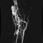
Location,
length, and enhancement: systematic approach to differentiating intramedullary spinal cord lesions. Arteriovenous malformation. A 16-year-old male with left leg weakness and progressive areflexia. a Sagittal T2 fat-saturated image demonstrates a conglomeration of flow voids within the cord parenchyma with surrounding hyperintensity. Prominent extramedullary flow voids lead to this conglomeration of vessels (arrow). b The sagittal T1 post-contrast image shows a nodule of enhancement (arrowhead). c Conventional angiogram shows an artery feeding a nidus (arrow), which is drained by a prominent vein

This image
is part of a series which can be scrolled interactively with the mousewheel or mouse dragging. This is done by using Template:Imagestack. The series is found in the category Cerebral arteriovenous malformation MRT TOF MIP Case 001. Arteriovenöse Malformation im Gehirn: MRT TOF gedrehte MIP-Projektion


Arteriovenöse
Malformation des Gehirns in der Computertomographie nativ, also ohne Kontrastmittel. Die erweiterten Gefäßkonvolute sind dennoch hyperdens also hell zu erkennen. Vergleiche auch die MRT-Bilder dieses Falls.

Arteriovenöse
Malformation des Gehirns in der Computertomographie nativ, also ohne Kontrastmittel. Die erweiterten Gefäßkonvolute sind dennoch hyperdens also hell zu erkennen. Vergleiche auch die MRT-Bilder dieses Falls.

Arteriovenöse
Malformation des Gehirns in der Computertomographie nativ, also ohne Kontrastmittel. Die erweiterten Gefäßkonvolute sind dennoch hyperdens also hell zu erkennen. Vergleiche auch die MRT-Bilder dieses Falls.

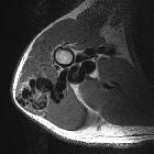
PTEN-Hamartom-Tumor-Syndrom:
Digitale Subtraktionsangiographie einer arteriovenösen Malformation am Knie bei PTEN-Hamartom-Tumor-Syndrom. Beachte die multiplen, kugelig wirkenden intranidalen „flow-related“ Aneurysmen.

Arteriovenous malformations (AVMs) are characterized by an abnormal leash of vessels allowing for arteriovenous shunting. They can occur anywhere in the body but have a predilection towards the head and neck . There is direct arteriovenous communication with no intervening capillary bed. They can be congenital or acquired .
Classification
Location-specific subtypes
- cerebral arteriovenous malformation
- hepatic arteriovenous malformation
- musculoskeletal arteriovenous malformation
- pulmonary arteriovenous malformation
- renal arteriovenous malformation
- uterine arteriovenous malformation
Siehe auch:
- arteriovenöse Malformationen der Lunge
- zerebrale arteriovenöse Malformation
- Spetzler AVM grading system
- arteriovenöse Malformation des Uterus
- PTEN-Hamartom-Tumor-Syndrom
- Arteriovenöse Malformation des Gesichts
- F.P. Weber-Syndrom
und weiter:
- Developmental Venous Anomaly
- kavernöse Transformation Pfortader
- Neurofibromatose Typ 1
- intraventrikuläre Blutung
- Intrakranielle Blutung
- durale AV-Fistel
- Balkenläsionen
- Hämangioblastom
- Ponsblutung
- solitärer pulmonaler Rundherd
- Exophthalmus
- neuroradiologisches Curriculum
- Superfizielle Hämosiderose der Leptomeningen
- Wyburn-Mason syndrome
- FCE
- macrodystrophia lipomatosa
- arteriovenöser Shunt Leber
- yasargil classification
- Tumor im Corpus callosum
- kongenitale vaskuläre Malformationen
- Gefäßverkalkungen
- intrakranielle vaskuläre Malformationen
- premature closure of a growth plate
- Temporallappenepilepsie
- Vena Galeni Malformation
- Spasmus hemifacialis
- spinale arteriovenöse Malformationen
- cerebral malformation
- intrahepatic arterio-portal shunt
- Spetzler-Martin AVM grading system
- Arteriovenöse Malformation der Niere
- fetal arteriovenous malformations
- SOLAMEN-Syndrom
- low flow vascular malformation
- renal cell carcinoma with an arteriovenous malformation
- intrakranielle Arteriosklerose
- arteriovenous malformation of the hand
- Gefäßmalformation Niere
- vaskuläre intraorbitale Läsionen
- paravertebrale arteriovenöse Malformation
- arteriovenöse Malformation der Orbita


 Assoziationen und Differentialdiagnosen zu Arteriovenöse Malformation:


