Cephalocele


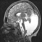
Cephalocele refers to the outward herniation of CNS contents through a defect in the cranium. The vast majority are midline.
Epidemiology
The estimated incidence is 0.8-4:10,000 live births with a well recognized geographical variation between types; however, this has been speculated to be underestimated as many may result in elective termination, in utero demise, or stillbirth. There may be a female predilection .
Clinical presentation
The presentation may vary widely depending upon the type of defect.
Examples include:
- large mass seen extending from cranium on prenatal ultrasound
- small cranial soft tissue mass palpated in childhood (e.g. atretic cephalocele)
- in utero demise (severe defect)
Pathology
It is thought to arise due to failed closure of the rostral end of the neuropore. This may result from either overgrowth of neural tissue in the line of closure, or failure of induction by adjacent mesodermal tissues which interferes with normal skull closure.
Classification
Cephaloceles can be classified into 5 types, based upon the herniated contents :
- meningocele: CSF lined by meninges
- gliocele: CSF lined by glial tissue
- meningoencephalocele: CSF, brain, and meninges
- meningoencephalocystocele: CSF, brain, meninges, and part of a ventricle and choroid plexus
- atretic cephalocele: dura, fibrous tissue, and degenerated brain
Location
- occipital cephalocele: most common, up to 75%
- parietal cephalocele: up to 37%
- frontal cephalocele/fronto-ethmoidal cephalocele: ~10%, this type is most common in Asia
- petrous apex cephalocele : rare
- intra sphenoidal cephalocele : rare
Associations
Additional congenital anomalies may be present in up to 50 % of cases. They include
- aneuploidic
- non-aneuploidic syndromic
- non-syndromic CNS anomalies
- fetal hydrocephalus/neonatal hydrocephalus
- microcephaly: ~20% of cases
- Dandy-Walker continuum
- dysgenesis of corpus callosum
- spina bifida
- non-syndromic non-CNS anomalies
- cleft lip and palate
- congenital cardiovascular anomalies
- hypertelorism
- amniotic band syndrome : if there is an unlucky slash defect around the occipital region
Markers
- maternal serum alpha-fetoprotein (MSAFP) may be elevated
Radiographic features
Ultrasound
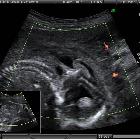
Sonographically, these lesions may appear as:
- a cyst protruding from the fetal calvarium representing a meningocele or cyst within cyst appearance
- a solid mass protruding from the calvarium representing a herniated brain: encephalocele
- either or both of the above associated with a defect in the calvarium
Treatment and prognosis
The overall prognosis is variable dependent on severity and other associations (presence of hydrocephalus, microcephaly, etc). If a large cephalocele is noted in an antenatal ultrasound scan, it generally implies a poor prognosis.
Siehe auch:
- Spina bifida
- Pätau-Syndrom
- Amniotisches-Band-Syndrom
- Herzfehler
- Meckel-Syndrom
- Hypertelorismus
- Meningozele
- Trisomie 18
- Mikrozephalie
- Lippen-Kiefer-Gaumen-Spalte
- Meningoenzephalozele
- Walker-Warburg-Syndrom
- Dandy Walker continuum
- Frontonasale Dysplasie
- kongenitaler Hydrozephalus
- atretische parietale Cephalocele
- dysgenesis of corpus callosum
und weiter:

 Assoziationen und Differentialdiagnosen zu Zephalozele:


