cerebral vascular territories


An understanding of brain arterial vascular territories is important in understanding stroke and complications from surgery and endovascular procedures.
Although one could be excused for thinking that within the brain, such a carefully organized organ, blood supply would be constant, the truth is that a great deal of variety exists. Two main factors contribute to vascular territories:
As a result, different sources will have surprisingly different diagrams and descriptions. What is presented here is a general ball-park scheme, with a brief description of the main branches and anatomy.
It is beyond the scope of this single article to describe each vessel/branch in detail: refer to the relevant subarticle if required.
General overview
The intracranial circulation can be conveniently divided into anterior and posterior circulation, on the basis of internal carotid artery and vertebral artery supply respectively.
- anterior circulation
- posterior circulation
Anterior cerebral artery
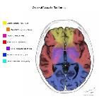
The anterior cerebral artery (depicted in yellow on the diagrams) is a terminal branch of the internal carotid artery. It may be divided into 2 or 3 segments, depending on the author.
A1 segment: from origin to anterior communicating artery and gives rise to medial lenticulostriate arteries (inferior parts of the head of the caudate and the anterior limb of the internal capsule)
- A2 segment: from anterior communicating artery to bifurcation in pericallosal artery and callosomarginal artery
- A3 segment: major branches, excluding terminal branches, which supply the medial portions of frontal lobes, superior medial part of parietal lobes, anterior part of the corpus callosum
Medial lenticulostriate arteries
Branches of the A1-segment of the anterior cerebral artery. They supply the anterior inferior parts of the basal nuclei and the anterior limb of the internal capsule. Heubner's artery is the largest of the medial lenticulostriate arteries and supplies the anteromedial part of the head of the caudate and anterior inferior internal capsule.
Middle cerebral artery
The cortical branches of the MCA (depicted in red in the diagrams) supply the lateral surface of the hemisphere, except for the medial part of the frontal and the parietal lobe, which is supplied by the ACA, and the inferior part of the temporal lobe, which is supplied by the PCA.
Lateral lenticulostriate arteries
The deep penetrating branches are called the lateral lenticulostriate arteries. The territory of the lateral lenticulostriate perforating arteries of the MCA is indicated with a different color (purple) from the rest of the territory of the MCA because it is a well-defined area supplied by penetrating branches, which may be involved or spared in infarcts separately from the main cortical territory of the MCA.
Posterior cerebral artery
The vascular territory of the PCA is depicted in blue. The P1 segment extends from the PCA origin to the posterior communicating artery, contributing to the circle of Willis. Posterior thalamoperforating arteries branch off the P1 segment to supply blood to the midbrain and thalamus. Cortical branches of the PCA supply the inferomedial part of the temporal lobe, occipital pole, visual cortex, and splenium of the corpus callosum. In addition, the arterial supply of hippocampus usually arises from PCA, including:
- anterior hippocampal artery, which usually arises from the PCA and less commonly from the anterior choroidal artery
- larger middle hippocampal artery, most commonly arising from the PCA
- posterior hippocampal artery, usually arising from the splenial artery or the PCA
Anterior Choroidal artery (AchA)
The territory of the AChA is part of the hippocampus, the posterior limb of the internal capsule and extends upwards to an area lateral to the posterior part of the cella media.
Watershed zones
Watershed infarcts occur at the border zones between major cerebral arterial territories as a result of hypoperfusion. There are two patterns of border zone infarcts:
Infarctions of the cortex and adjacent subcortical white matter located at the border zone of ACA/MCA and MCA/PCA
Infarctions of the deep white matter of the centrum semiovale and corona radiata at the border zone between lenticulostriate perforators and the deep penetrating cortical branches of the MCA or at the border zone of deep white matter branches of the MCA and the ACA
Corpus callosum
The corpus callosum has a rich blood supply, relatively constant and is uncommonly involved by infarcts. The majority of the corpus callosum (CC) is supplied by the pericallosal arteries and the posterior pericallosal arteries, branches from the anterior and posterior cerebral respectively. In 80% of patients additional supply comes from the anterior communicating artery, via either subcallosal artery or median callosal artery.
- the subcallosal artery (50% of patients) is essentially a large version of a hypothalamic branch, which in addition to supplying part of the hypothalamus also supplies the medial portions of the rostrum and genu.
- the median callosal artery (30% of patients) can be thought of as a more extended version of the subcallosal artery, in that it travels along the same course, supplies the same structures but additionally reaches the body of the corpus callosum.
- the posterior pericallosal artery (also known as splenial artery) supplies a variable portion of the splenium. Its origin is inconstant, arising from P3 or branches thereof.
Basal Ganglia
The basal ganglia are supplied by perforating branches of the anterior cerebral artery and middle cerebral artery, mainly the medial lenticulostriate arteries and lateral lenticulostriate arteries.
- Medial lenticulostriate arteries, from the anterior cerebral artery, supply the globus pallidus and medial portion of the putamen.
- Lateral lenticulostriate arteries, from the middle cerebral artery, supply the lateral portion of the putamen.
- Recurrent artery of Heubner, arising as a large branch from the anterior cerebral artery, supplies the caudate nucleus.
Brainstem
The brainstem is supplied by the vertebrobasilar circulation and their branches, small branches of the posterior cerebral artery (PCA), and the anterior spinal artery.
Small penetrating branches from:
- medial branches of the superior cerebellar artery
- pontine branches of basilar artery, thalamoperforator arteries
- posterior inferior cerebellar artery (PICA)
- anterior spinal artery
- direct branches of the distal vertebral artery
Cerebellum
The cerebellum is essentially supplied by three vessels:
Superior cerebellar artery (SCA)
This vessel supplies:
- whole superior surface of the cerebellar hemispheres down to the great horizontal fissure
- the superior vermis
- dentate nucleus
- most of the cerebellar white matter
Anterior inferior cerebellar (AICA)
The amount of tissue supplied by the AICA is variable (PICA-AICA dominance) but usually includes:
- middle cerebellar peduncle
- inferolateral portion of the pons
- flocculus
- anteroinferior surface of the cerebellum
Posterior inferior cerebellar (PICA)
Has a variable territory depending on the size of the AICA, but usually supplies:
- posteroinferior cerebellar hemispheres (up to the great horizontal fissure)
- inferior portion of the vermis
- 18% arise extracranially, inferior to the foramen magnum
- 10% arise from the basilar rather than vertebral artery
- 2% bilaterally absent
- occasionally loops around the cerebellar tonsil
It divides into lateral and medial branches that supply the inferior portion of the vermis and cerebellar hemispheres respectively.
Direct basilar/vertebral arterial branches
These branches supply the medulla oblongata and the pons.
NOTE: occasionally a small vertebral artery will terminate into a common PICA/AICA trunk.


 Assoziationen und Differentialdiagnosen zu arterielle Versorgungsgebiete des Gehirns: