Hüftkopfnekrose


Avascular necrosis of the hip is more common than other sites, presumably due to a combination of precarious blood supply and high loading when standing.
Clinical presentation
The most common presenting symptom is a pain in the region of affected hip, thigh, groin, and buttock. Although few patients may remain asymptomatic until late stages.
Pathology
Typically it affects the superior articular surface (between 10-2 o'clock) and begins in the most anterior part of the hip.
Etiology
It can be thought of as traumatic (secondary to the neck of femur fractures) or non-traumatic. In non-traumatic cases, it is bilateral in 40%.
- traumatic
- chronic corticosteroid therapy
- alcoholism
- smoking
- systemic lupus erythematosus (SLE)
- hyperlipidaemias
- HIV
- hemoglobinopathies
- chronic renal failure
- diabetes mellitus
- pregnancy-related
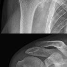
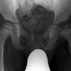
Radiographic features
Specific staging system (Ficat staging) exists for the hip which includes x-ray, MRI and bone scan appearances, and covers much of the imaging appearances, thus please refer to that article.
Other than describing the general appearance of the affected region, the following are necessary to include in the report as they have a bearing on prognosis and treatment:
- position
- estimating percentage volume of the head involved (axial) and percentage weight-bearing surface involved (coronal)
- coexisting osteoarthritis or secondary degenerative change
- joint effusion
- presence of a potentially unstable osteochondral fragment: rim sign
- subchondral fractures
CT
Often more sensitive than plain film in showing subchondral fractures.
MRI
MRI is the most sensitive modality, with a sensitivity of 71-100% and specificity of 94-100%. As there is a high rate of bilateral involvement, both hips should be included in the field of view of at least some sequences.
- T1: usually the initial specific findings are areas of low signal representing edema, which can be bordered by a hyperintense line which represents blood products
- T2: may show a second hyperintense inner line between normal marrow and ischemic marrow. This appearance is highly specific for AVN hip and known as "double line sign".
The Mitchell classification is commonly used to classify AVN based on MR-images.
Differential diagnosis
In some situations consider
- subchondral insufficiency fracture of the femoral head - considered by some as a different entity
General imaging differential considerations include:
- hematopoietic marrow (see bone marrow)
- Pitt's pit
- fovea centralis
- idiopathic transient osteoporosis of the hip (ITOH)
- hyperemia with diffuse increased uptake of radiotracer by the femoral head, neck, and intertrochanteric region
- chondroblastoma
- fracture
- infection
- pain and fever
- usually involves both sides of the joint
- metastases
See also
- avascular necrosis - general article
- Legg-Calve-Perthes disease
- Ficat and Arlet staging
- Steinberg staging of avascular necrosis
- Mitchell classification of avascular necrosis
- ARCO classification of osteonecrosis
Siehe auch:
- Osteomyelitis
- Aseptische Knochennekrose
- synovial herniation pit
- Fibröse Dysplasie
- Arthrose
- Epiphyseolysis capitis femoris
- Chondroblastom
- Schenkelhalsfraktur
- idiopathische kindliche Hüftkopfnekrose
- Knochenmark
- Hämophilie
- Ficat staging
- transiente Osteoporose der Hüfte
- Meyersche Dysplasie
- Rapidly destructive coxarthrosis
- rim sign
- Herring-Zeichen
- Klassifikation Morbus Perthes
und weiter:


 Assoziationen und Differentialdiagnosen zu Hüftkopfnekrose: