ossifying fibroma


Ossifying fibromas are benign bone lesions that should be differentiated from non-ossifying fibromas and fibrous dysplasia. Osteofibrous dysplasia is considered as a separate pathological entity in view of its different presentation and treatment, although histopathologically similar to ossifying fibroma.
Epidemiology
These lesions are most frequently encountered in young children (often <10 years).
Pathology
Histology
They comprise haphazardly distributed lamellated bony spicules on a background of fibrous stroma. Despite being benign, they can be locally aggressive. Immunohistochemical staining of lesions shows positive keratin cells in the majority of the cases.
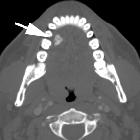
Location
- lower extremity
- tibia: most frequent site (90% of the time); there is a predilection for the anterior tibial cortex
- femur: occurs in a diaphysial location
- mandible and maxilla: these are examples of cementum-poor cemento-ossifying fibromas (see WHO classification scheme for odontogenic tumors)
- sinonasal: expansile lesions with peripheral ossification and central lucency
Associations
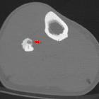
Radiographic features
Plain radiograph and CT
- well-circumscribed lesion
- evidence of intracortical osteolysis with a characteristic sclerotic band (osteoblastic rimming)
- moderate cortical expansion
- homogeneous lesion matrix
MRI
Reported signal characteristics include
- T1: low signal
- T2: iso-high signal
- T1 C+ (Gd): typically shows enhancement
Treatment and prognosis
Tend to regress over time. For locally aggressive lesions, surgical resection is often curative although recurrence has been reported.
Complications
Differential diagnosis
Imaging differential considerations include
- fibrous dysplasia: has no osteoblastic rimming
- adamantinoma: may share a common origin with ossifying fibromas
- osteoid osteoma
- osteofibrous dysplasia
Siehe auch:
- Osteoid-Osteom
- Fibröse Dysplasie
- nicht ossifizierendes Fibrom
- Kryptorchismus
- Jaffé-Campanacci-Syndrom
- Adamantinom der langen Röhrenknochen
und weiter:
- Zementoblastom
- radiologisches muskuloskelettales Curriculum
- Knochentumoren
- arrested pneumatization of skull base
- fibröser Kortikalisdefekt
- differential diagnosis for calcified masses in the mandible
- arrested pneumatisation of the skull base
- Stein der Nasennebenhöhlen
- ossifying fibroma of the mandible
- ossifizierendes Fibrom des Schädels
- knochenbildende Tumoren


 Assoziationen und Differentialdiagnosen zu Ossifizierendes Fibrom: