pilomatricoma
A pilomatricoma is an uncommon, benign neoplasm thought to arise from hair cortex cells. It was formerly referred to as pilomatrixoma or calcifying epithelioma of Malherbe.
Epidemiology
- the reported incidence ranges between 1 in 500-2000
- they make up 0.12% of cutaneous neoplasms and 20% of all hair follicle related tumors
- more common in females; F:M ratio = 3:2
- more common in the Caucasian population
- more common in children, but occurrence in adults is increasingly being recognized
- there is a developmental bi-modal peak during the first and sixth decades of life
- usually develops within the first two decades of life with 40% of cases occurring before the age of 10 years and 60% before the age of 20 years
- usually solitary but up to 3% are multiple
Clinical presentation
- painless
- slow-growing
- superficial
- mobile
- hard mass
- ranges in size from 0.5 to 5 cm in diameter
- overlying skin has a bluish-red discolouration
Pathology
Location
Approximately 50% of cases occur in the head and neck region. This is followed by the upper extremities, trunk and the lower extremities. Less commonly seen in the frontal, temporal, cheek, periorbital and preauricular areas.
Macroscopic appearance
Well-circumscribed nodulocystic tan colored mass containing irregularly shaped, lobulated islands of cells separated by fine, fibrovascular connective tissue stroma.
Microscopic appearance
- peripheral basaloid cells are darkly stained, round or elongated, with indistinct cell borders and minimal cytoplasm; they contain round to ovoid deeply basophilic nuclei with most showing prominent nucleoli
- central ghost cells have abundant, pale, eosinophilic cytoplasm with well defined cell borders and central clear area
Associations
Recognized associations include:
- sarcoidosis
- Turner syndrome
- Gardner syndrome
- Steinert disease
- Rubinstein-Taybi syndrome
- myotonic muscular dystrophy (associated with multiple and recurring lesions)
- skull dysostosis
Familial forms may be present.
Variants
Pilomatrix carcinoma = malignant pilomatricoma (rare)
Radiographic features
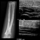
Plain radiograph
- there may be non-specific soft tissue calcification
Ultrasound
- ovoid complex mass, often with calcification, at the junction of the dermis and subcutaneous fat with focal thinning of the overlying dermis
- appear as target lesions with a hyporeflective connective tissue capsule and central hyper-reflective epithelial cells
CT
- well-defined, subcutaneous tumors with microcalcifications
MRI
- T1: uniform, homogeneously low to intermediate to high signal
- T2: variable appearance either inhomogeneous with multiple areas of intermediate signal or homogeneous with either intermediate signal intensity or high signal radiating from the center
- T1 C+ (Gd): variable post contrast T1 appearance, where some only enhance peripherally
Treatment and prognosis
Surgical resection is often curative.
History and etymology
It was first described by Malherbe and Chenantias in 1880, as a subcutaneous tumor, and was shown to be a derivative from hair cortex cells in 1942 by Turhan and Krainer.
The root of the term pilomatricoma is derived from the Latin word pilus meaning hair.
Differential diagnosis
General imaging differential considerations include:
- epidermal inclusion cyst
- ossifying hematoma
- giant cell tumor
- chondroma
- dermoid cyst
- foreign body reaction
- degenerating fibroxanthoma
- metastatic bone formation
- osteoma cutis
Siehe auch:
und weiter:


 Assoziationen und Differentialdiagnosen zu Malherbe-Tumor:


