soft tissue calcification

Heterotope
Ossifikationen im Verlauf der ischiokruralen Muskulatur am ehesten als Restzustand einer älteren Teilruptur/Einblutung.

Radiological
identification and analysis of soft tissue musculoskeletal calcifications. Gout. Frontal radiograph of the right foot shows tophi and erosions. Some tophi may present with mineralisation (arrowheads)

Calcinosis
cutis bei kutaner Graft-versus-Host Reaktion nach Stammzelltransplantation: Die Computertomografie (links 3 Schichten axial, rechts Volumen Rendering mit unterschiedlicher Transparenz der Gewebe) zeigt die ausgeprägten flächigen Verkalkungen. Beachte auch die Verkalkung in der V. cava inferior, die als Residuum einer Cavathrombose zu deuten ist.

Radiological
identification and analysis of soft tissue musculoskeletal calcifications. Synovial osteochondromatosis. Anteroposterior radiograph of the left hip shows multiple intra-articular osteocartilaginous bodies (arrows) with ring-like calcifications centred on the coxofemoral joint

Radiological
identification and analysis of soft tissue musculoskeletal calcifications. Renal insufficiency with tumoural calcinosis. (a) Anteroposterior radiograph of the pelvis and (b) axial CT image of the left hip show tumoural calcinosis (arrow) around the left hip with a multiloculated appearance in a clinical context of chronic renal failure. Note fluid-fluid level with denser material layering more posteriorly (arrowhead in b). I: ischium, T: greater trochanter

Radiological
identification and analysis of soft tissue musculoskeletal calcifications. Cysticercosis. Anteroposterior radiograph of the pelvis shows multiple cigar-shaped calcifications characteristic of cysticercosis

Radiological
identification and analysis of soft tissue musculoskeletal calcifications. Phleboliths. Anteroposterior radiograph of the elbow shows phleboliths in the soft tissues with the characteristic central lucency (arrowhead) in a patient with a venous malformation of the upper extremity. Note multifocal soft tissue masse effect that corresponds to the diffuse venous malformation (arrows)

Calcinosis
universalis • Dermatomyositis - Ganzer Fall bei Radiopaedia

Radiological
identification and analysis of soft tissue musculoskeletal calcifications. Vascular calcifications. (a) Oblique radiograph of the femur shows atherosclerotic vascular calcifications (arrowheads) and (b) lateral radiograph of the ankle shows metabolic vascular calcifications (arrows)

Radiological
identification and analysis of soft tissue musculoskeletal calcifications. Systemic Sclerosis. Posteroanterior radiograph of the hand shows tumoural calcinosis in the soft tissue (arrow). Note acro-osteolysis (arrowhead) and atrophy of soft tissue (curved arrow), characteristic of systemic sclerosis

Radiological
identification and analysis of soft tissue musculoskeletal calcifications. Periarticular calcifications following corticoid injection. (a) Frontal, (b) oblique and (c) lateral radiographs of the wrist show palmar soft tissue calcifications (arrowhead) following carpal tunnel injection of corticoid

Radiological
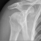
identification and analysis of soft tissue musculoskeletal calcifications. HADD. (a) Anteroposterior radiograph of the right shoulder shows a calcific tendinopathy of the supraspinatus tendon (arrowhead) during formative or resting phase. (b) Anteroposterior radiograph of the right shoulder in a different patient shows migration of the calcifications to the subdeltoid-subacromial bursa (arrows) during the resorptive phase

Radiological
identification and analysis of soft tissue musculoskeletal calcifications. Schwannoma. (a) axial CT image of the sacrum shows calcifications in a lesion and (b) sagittal T2-weighted MRI image of the sacrum (arrows) and axial fat-saturated T1-weighted MRI image of the sacrum following gadolinium enhancement show the specific signal inside the schwannoma (arrowheads)

Radiological
identification and analysis of soft tissue musculoskeletal calcifications. Disc calcifications. Sagittal CT images of the lumbar spine shows (a) calcium pyrophosphate dehydrate (CPPD) crystal deposition (arrowheads in a), (b) hydroxyapatite crystal deposition (arrowhead in b) and (c) syndesmophytes (arrowhead, in c) in three different patients. Note calcifications in ligamentum flavum and interspinous ligament associated with CPPD deposition disease (arrows in a) and ossification of disc space associated with ankylosing spondylitis (arrow in c). Note linear calcifications paralleling the vertebral endplates in b, associated with height loss, corresponding to subacute fractures in a patient with osteoporosis

Radiological
identification and analysis of soft tissue musculoskeletal calcifications. Rapid degenerative arthropathy. (a) Anteroposterior radiographs initially and (b) 3 months later show progressive bone lysis (arrows) and periarticular calcifications (arrowheads)

Radiological
identification and analysis of soft tissue musculoskeletal calcifications. Chondrocalcinosis in the knee. (a) Anteroposterior radiograph of the left knee and (b) coronal fat-saturated proton density (PD)-weighted MRI image show chondrocalcinosis in the menisci (arrowheads)

Hand
radiograph showing tumoral calcinosis (metastatic calcification) in a patient on dialysis.

Soft tissue
calcification • Dermatomyositis - Ganzer Fall bei Radiopaedia

Oblique hand
radiograph showing tumoral calcinosis (metastatic calcification) in a patient on dialysis.

Soft tissue
calcification • Hyperparathyroidism - Ganzer Fall bei Radiopaedia

Radiological
identification and analysis of soft tissue musculoskeletal calcifications. Cervical facet joint calcifications. (a) Axial and (b) sagittal CT images of the cervical spine show dense material (arrowheads) in the right C2–C3 facet joint. Note the associated bone erosion of the right lamina (arrow)

Radiological
identification and analysis of soft tissue musculoskeletal calcifications. Dermatomyositis. (a) Lateral and (b) frontal radiographs of the left leg show sheet-like muscular calcifications in a case of dermatomyositis

Radiological
identification and analysis of soft tissue musculoskeletal calcifications. Synovial sarcoma. (a) Frog-leg radiograph, (b) axial CT image and (c) axial T1-weighted image of the left thigh show faint calcifications (arrow in a, b and c) that are located inside a tumour which is better seen on the CT and MRI assessment (arrowhead in a, b and c). Synovial sarcoma was found at pathology

Radiological
identification and analysis of soft tissue musculoskeletal calcifications. Supraspinatus calcifying tendinopathy with intraosseous extension. (a) Frontal radiograph, (b) coronal reformat CT image and (c) coronal fat-saturated T2-weighted image show supraspinatus amorphous calcifications (arrows) with intraosseous extension causing erosions (arrowheads). Note the sclerosis around the erosion, hyperdense in (a) and (b) and hypointense in (c)

Soft tissue
calcification • Tumoral calcinosis - Ganzer Fall bei Radiopaedia

Soft tissue
calcification • Heterotopic ossification (post amputation) - Ganzer Fall bei Radiopaedia

Radiological
identification and analysis of soft tissue musculoskeletal calcifications. CPPD arthropathy of the wrist. Posteroanterior radiograph of the wrist shows joint space narrowing and dense subchondral sclerosis at the radioscaphoid and lunocapitate joints (arrows) associated with widening of the scapholunate space, consistent with a SLAC wrist. Note chondrocalcinosis at the ulnocarpal joint space within the triangular fibrocartilage (arrowhead)

Soft tissue
calcification • Soft tissue calcification from chronic venous insufficiency - Ganzer Fall bei Radiopaedia

Radiological
identification and analysis of soft tissue musculoskeletal calcifications. Ossification. Axial CT image of myositis ossificans in the infraspinatus muscle shows a cortical and trabecular mineralisation pattern with a zonal distribution (absence of mineralisation centrally due to fatty bone marrow)

Teenager hit
in the right thigh 1 month ago with a shot put. AP (left) and lateral (right) radiographs of the femur show an amorphous area of calcification anterior and lateral to the femur.The diagnosis was myositis ossificans.

Radiological
identification and analysis of soft tissue musculoskeletal calcifications. Calcifying tendinopathy of flexor carpi ulnaris. Oblique radiograph of the wrist shows HADD in the flexor carpi ulnaris with amorphous cloudlike calcifications (arrowhead)

Soft tissue
calcification • Myositis ossificans progressiva - Ganzer Fall bei Radiopaedia

Radiological
identification and analysis of soft tissue musculoskeletal calcifications. CPPD crystal deposition disease in the knee. Lateral radiograph of the right knee shows chondrocalcinosis in hyaline cartilage at the posterior femoral condyle (arrow), in the gastrocnemius proximal tendon (arrowhead) and in the synovial lining at the suprapatellar recess (curved arrow)

Radiological
identification and analysis of soft tissue musculoskeletal calcifications. Metastatic calcified lung adenocarcinoma. (a, b) Two axial CT images of the thorax show calcified lung adenocarcinoma (arrowhead) (a) and calcified soft tissue chest wall metastasis (arrow) (b)

Scleroderma
• Scleroderma - hand manifestations - Ganzer Fall bei Radiopaedia

Salt and
pepper sign (skull) • Hyperparathyroidism - Ganzer Fall bei Radiopaedia

Pelvic
heterotopic ossification: when CT comes to the aid of MR imaging. A 35-year-old male patient with leg contractures, affected by posterior element incomplete fusion (spina bifida occulta). a MR T1-weighted axial images evidence a soft tissue abscess (small arrow) of the perineal region related to pressure sores. Lesion is determining a certain retraction over the superficial layers of the dermis. b STIR images show the focal inflammatory lesion partially filled with fluid and a large bursitis of the right gluteal region (large arrow). c On coronal T1-weighted fat-sat images after gadolinium administration, the abscess walls appear highly vascularised. The gluteal bursitis seems to be communicating with the infectious process, suggesting a septic bursitis. A prominent contrast enhancement is also seen to the sacrum, suggesting a bony involvement by the septic process. d Axial CT imaging performed with patient in a different leg positioning . The exam validates the connection between the perineal abscess and the gluteal bursitis. Amorphous calcifications (asterisks) are enclosed within the septic bursitis at the right gluteal region


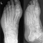
Gout in a
radiograph of the [left] foot. Typical (main) localization at the metatarsophalangeal joint of the great toe. Note also the soft tissue swelling on the lateral border of the foot.

Calcific
myonecrosis following snake bite: a case report and review of the literature. Initial anterior-posterior and lateral plain radiographs of left leg.
Soft tissue calcification is commonly seen and caused by a wide range of pathology.
Differential diagnosis
There is a wide range of causes of soft tissue calcification :
- dystrophic soft tissue calcification (most common)
- vascular
- arterial calcification
- phleboliths
- metabolic
- CPPD (causing chondrocalcinosis)
- metastatic calcification
- idiopathic tumoral calcinosis
- autoimmune diseases
- infection
- neoplasm
- primary or metastatic osteosarcoma
- synovial osteochondromatosis
- trauma
- myositis ossificans
- tendonitis
- injection site granuloma
- surgical scar
- burn
- others
Siehe auch:
- synoviale Osteochondromatose
- Osteosarkom
- Gicht
- Myositis ossificans
- Tendinitis calcarea
- primärer Hyperparathyreoidismus
- tumoröse Kalzinose
- Synovialsarkom
- Hydroxylapatit-Ablagerungserkrankung
- Heterotope Ossifikation
- Chondrokalzinose
- calcinosis circumscripta
- Kalziumpyrophosphat-Ablagerungskrankheit
- Kristallarthropathie
- chronische Niereninsuffizienz
- Burnett-Syndrom
- dystrophic soft-tissue calcification
- intramuskuläre Verkalkungen
- Hypervitaminose D
- Calcinosis cutis universalis
- verkalkende Myonekrose
- muskuloskelettale Manifestationen Dermatomyositis
- Weichteilverkalkungen bei chronisch venöser Insuffizienz
- Neuritis ossificans
- Merkspruch Weichteilverkalkungen
- Graft-versus-Host-Reaktion kutan
und weiter:
- periartikuläre Verkalkungen
- periprothetische Ossifikationen
- chondrocalcinosis (mnemonic)
- Pseudohypoparathyreoidismus
- generalized increased bone density in paediatrics
- calcium pyrophosphate dihydrate deposition arthropathy
- Calcinosis cutis
- Weichteilverkalkungen der Hand
- Synovialsarkom mit Verkalkungen
- juvenile Dermatomyositis
- verkalktes Hämatom
- Weichteilverkalkungen bei systemischem Lupus Erythematodes

 Assoziationen und Differentialdiagnosen zu Weichteilverkalkungen:
Weichteilverkalkungen
bei chronisch venöser Insuffizienz
Kalziumpyrophosphat-Ablagerungskrankheit


