temporal arteritis


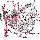
Giant cell arteritis (GCA) is a common granulomatous vasculitis affecting medium- to large-sized arteries. It is also known as temporal arteritis or cranial arteritis, given its propensity to involve the extracranial external carotid artery branches such as the superficial temporal artery.
Epidemiology
Giant cell arteritis is the most common primary systemic vasculitis. It has an incidence of 200 per million persons per year . Typically affects older individuals with patients usually being older than 50, with a peak incidence between the ages of 70 and 80 . There is a recognized female predilection.
Clinical presentation
There are many possible clinical features that present in a subacute fashion :
- headache (most common) with or without scalp tenderness
- systemic symptoms (e.g. fever, fatigue, weight loss)
- jaw claudication
- transient vision loss (amaurosis fugax)
- permanent vision loss (e.g. due to anterior ischemic optic neuropathy, central retinal artery occlusion, ischemic stroke, etc.)
- in such cases, there may be accompanying Charles-Bonnet syndrome
- weak pulse over the affected arteries
- bruits on auscultation over the affected arteries
- clinical features of an aortic aneurysm or dissection if the aorta is involved
Pathology
It is histologically similar to other large vessel vasculitides (such as Takayasu arteritis) showing granulomatous inflammation of arteries with infiltration predominantly by histiocytes, lymphocytes, and multinucleated giant cells. The characteristic multinucleated giant cells are only found in ~50% of cases .
Areas of normal superficial temporal artery interspersed within inflamed sections of artery, known as skip lesions, results in false negatives in up to 8-28% of cases .
In a study of 285 patients with biopsy-proven giant cell arteritis there were 4 main histological patterns :
Location
Can potentially affect any medium to large-sized vessels, affecting the aorta (~20% of cases ) and its major branches, particularly the extracranial branches of the carotid artery .
Markers
- serum erythrocyte sedimentation rate (ESR): markedly raised
- serum C-reactive protein (CRP): often markedly raised
Associations
- polymyalgia rheumatica: seen in ~50% of cases
Complications
- thoracic aortic aneurysms (more commonly ascending aorta)
- aortic dissection (more commonly ascending aorta)
Radiographic features
Ultrasound
- increased diameter of the superficial temporal artery and hypoechoic wall thickening (halo sign)
- with duplex ultrasound, sensitivity is 87% and specificity is 96%
- more specific for giant cell arteritis if bilateral
- reversible under corticosteroid treatment; this is reflected in the normalization of the sonographic features
- stenosis may be present but is not a specific sign for giant cell arteritis
CT
Findings on CT include:
- wall thickening of affected segments
- calcification
- mural thrombi
Arterial phase CT (angiography) is useful for assessing luminal abnormalities:
- stenoses
- occlusions
- dilatations
- aneurysm formation
MRI
- T1 C+ (Gd)
- the best sequence for assessment
- shows mural inflammation very well
- mean wall thickness increased in the affected region
- luminal diameter correspondingly decreased in the affected region
- reported approximate sensitivity and specificity is 80% and 97%, respectively
Treatment and prognosis
Treatment is with corticosteroid therapy and aspirin . Methotrexate may be used in combination with corticosteroid therapy initially, or as a corticosteroid-sparing drug .
Differential diagnosis
Imaging differential considerations include:
- Takayasu arteritis: affects younger patients (<50 years) and affects more proximal vessels
- atherosclerotic disease
Siehe auch:
und weiter:

 Assoziationen und Differentialdiagnosen zu Riesenzellarteriitis:


