Aortitis

Insights into
imaging of aortitis. a–d A 51-year-old male with chest pain. Spin-echo T1-weighted (a), post-gadolinium enhanced (b) and proton density (c) images of the aortic arch show diffuse and relatively uniform thickening of the aortic arch with focal aneurysmal dilatation of the posterior aspect of the distal arch (arrow). Vivid gadolinium enhancement is uniform and diffuse (b), being even more vivid at the mid-outer portion of the aortic wall (d), corresponding to a “double ring” appearance

Insights into
imaging of aortitis. a–c A 59-year-old woman with low-grade fever, malaise and high white blood cell count. Axial (a) CTA image demonstrates normal thickness (arrowhead) of the ascending aortic wall with minimal thickening of the descending aortic wall (arrow). Axial contrast-enhanced MR image (b) shows contrast in the ascending and descending aortic wall corresponding to vivid 18-FDG uptake on PET-CT image (c)

Syphilitic
aortitis. Thoracic Angio-CT (axial section on mediastinal window) shows irregular aortic wall thickening with soft-tissue accumulation owing to the chronic inflammatory process.

Insights into
imaging of aortitis. A 36-year-old male with abdominal pain and weight loss over the last 3 months. MDCTA of the abdomen demonstrates diffuse thickening of the aorta and superior mesenteric artery (SMA) with substantial narrowing of the SMA lumen (arrow). Courtesy of Dr. Ludmila Guralnik, Rambam Health Care Campus, Haifa, Israel

Aortitis as a
cause of acute coronary syndrome. MPR of the left main coronary ostium with severe stenosis due to aortic wall thickening (arrow).

Aortitis as a
cause of acute coronary syndrome. MIP CT with mural thickening of the aortic root, narrowed coronary ostia (back arows), normal aortic arch main branches (white arrowheads) and normal remainder of the aorta (white arrows).

Aortitis as a
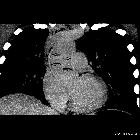
cause of acute coronary syndrome. Delayed enhancement CT coronal MPR of the ascending thoracic aorta with enhancement of the media and adventitia layers (arows).

Aortitis as a
cause of acute coronary syndrome. Delayed enhancement CT with diffuse swollen intima and enhanced media and adventitia (arrows).

Aortitis as a
cause of acute coronary syndrome. CT pre contrast with diffuse mural thickening and increased density of ascending aorta walls (arrows).

Insights into
imaging of aortitis. A 67-year-old female several months after ascending aneurysm repair, with low-grade fever and malaise. Axial non-contrast-enhanced (a) and contrast-enhanced (b) images of the chest demonstrate an ascending aortic graft with surrounding minimal aortic wall thickening (arrowheads in a and b respectively). The area was shown to be positive for moderate 18-FDG uptake on the PET-CT image (arrowhead in c). Blood cultures revealed Staphylococcus aureus infection, concerning for infectious aortitis

Insights into
imaging of aortitis. A 75-year-old male with abdominal pain. An axial non-contrast-enhanced CT image (a) of the abdomen demonstrates circumferential homogeneous thickening of the aortic wall up to 5 mm corresponding to vivid circumferential 18-FDG uptake on PET-CT image (b). Subsequent biopsy of the aortic wall was consistent with idiopathic inflammation

Insights into
imaging of aortitis. a–d A 48-year-old woman with weight loss, malaise and headaches. Axial (a, b) CTA images demonstrate concentric thickening of the brachiocephalic artery, continuing toward its branches: right common carotid artery, right subclavian artery and left subclavian artery (originating from the brachiocephalic artery in this case). Multiplanar curved reformats (c, d) demonstrate both wall thickening and aneurysmal dilatations along the course of both common carotid arteries (cright, dleft) and brachiocephalic artery trunk (c). Courtesy of Dr. Ludmila Guralnik, Rambam Health Care Campus, Haifa, Israel

Syphilitic
aortitis. Thoracic Angio-CT (axial section on mediastinal window) revealed aneurysmatic dilatation of the ascending aorta and the proximal thoracic descending aorta associated with a mural thrombus at the posterior wall.

Insights into
imaging of aortitis. a–c 20-year-old previously healthy woman with chest pain. CTA of the lower neck demonstrates circumferential wall thickening of the left carotid artery (a). Ascending aorta is dilated with circumferential wall thickening (b). Gadolinium-enhanced sagittal (c) view demonstrated dilatation of both ascending and descending thoracic aorta as well as aortic wall thickening including in the abdominal aorta

Insights into
imaging of aortitis. a–c A 25-year-old previously healthy man with chest and abdominal pain, weight loss and low grade fever. Axial (a), sagittal (b) and coronal (b) images of CTA of the torso demonstrate circumferential wall thickening of both the ascending and descending thoracic aorta as well as aortic wall thickening including in the abdominal aorta (white arrows). Diffuse narrowing of the aorta can be appreciated on all three views. Courtesy of Dr. Eduard Ghersin, Jackson Memorial Hospital, Miami, Florida

Mycotic
aneurysm • Infectious aortitis with mycotic pseudoaneurysm - Ganzer Fall bei Radiopaedia

Takayasu
arteritis • Takayasu arteritis - Ganzer Fall bei Radiopaedia

Mycotic
aneurysm • Fungal aortitis with mycotic aneurysms - Ganzer Fall bei Radiopaedia

Giant cell
arteritis • Giant cell aortitis - Ganzer Fall bei Radiopaedia

Behçet
disease • Behcet disease - aortitis - Ganzer Fall bei Radiopaedia

Aortitis with
extrinsic compression of left main trunk: role of cardiac computed tomography. Periaortic thickening that compressed the left main coronary artery.

Aortitis with
extrinsic compression of left main trunk: role of cardiac computed tomography. Cardiac CT revealed a periaortic thickening.

Aortitis as a
cause of acute coronary syndrome. CT MPR with RCA significant ostial stenosis due to aortic walls thickening (arrow).
Aortitis refers to a general descriptor that involves a broad category of infectious and non-infectious conditions where there is inflammation (i.e. vasculitis) of the aortic wall.
Clinical presentation
The presentation is non-specific with fever, pain and weight loss.
Pathology
Etiology
- infectious
- syphilitic aortitis
- tuberculous aortitis
- pyogenic aortitis: especially Salmonella infection
- aortitis due to HIV
- infected (mycotic) aortic aneurysm
- non-infectious
- giant cell arteritis
- Takayasu arteritis
- other rheumatologic disorders
- rheumatoid arthritis
- systemic lupus erythematosus
- granulomatosis with polyangiitis
- Behçet disease
- polyarteritis nodosa
- microscopic polyangiitis
- HLA-B27–associated seronegative spondyloarthropathies
- Cogan syndrome
- chronic periaortitis
- idiopathic aortitis
- radiation-induced aortitis
- IgG4-related cardiovascular disease
Siehe auch:
- Morbus Bechterew
- Polyarteriitis nodosa
- Rheumatoide Arthritis
- Vaskulitis
- Granulomatose mit Polyangiitis
- systemischer Lupus Erythematodes
- Morbus Behçet
- Takayasu-Arteriitis
- Riesenzellarteriitis
- Mikroskopische Polyangiitis
- mykotisches Aortenaneurysma
- syphilitische Aortitis
- Reaktive Arthritis
- tuberculous aortitis
- infektiöse Aortitis
- Großgefäßvaskulitis
- radiation induced aortitis
- pyogenic aortitis
- aortitis due to HIV
- idiopathic aortitis
- Cogan-Syndrom
- granulomatöse Aortitis
- chronische Periaortitis
und weiter:

 Assoziationen und Differentialdiagnosen zu Aortitis:


