tuberculous osteomyelitis


Tuberculous osteomyelitis is one of the rarer musculoskeletal manifestations of tuberculosis.
Epidemiology
Tuberculous osteomyelitis accounts for ~20% of musculoskeletal tuberculosis .
Clinical presentation
Patients may present with a painful "cold abscess" with a localized mass/swelling +/- draining sinus with erythema or warmth; a low-grade fever may be present .
There is often a delay between presentation and diagnosis, with a median time to diagnosis reported as 26.4 months .
Pathology
Most cases are caused by Mycobacterium tuberculosis with non-tuberculosis mycobacterial infections very rare although increased in the settings of AIDS .
Location
Isolated tuberculosis osteomyelitis without associated tuberculous arthropathy most commonly occurs in the metaphyses of the
- femur
- tibia
- small bones of the hand and foot (tuberculous dactylitis)
Radiographic features
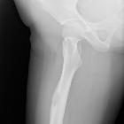
Plain radiograph
Plain radiographs can be normal in early infection and when abnormal can show :
- eccentric lytic lesion with minimal or no periosteal reaction
- a cortical defect may be present
- local osteopaenia
MRI
Tuberculous osteomyelitis has a variable appearance with signal characteristics similar to pyogenic osteomyelitis (i.e. low T1, high T2) being reported. One study of 11 cases has shown that some cases may have a slightly higher T1 peripheral rim and low-to-intermediate T2 signal and association with soft tissue abscess .
Differential diagnosis
- chronic pyogenic osteomyelitis: in the skeletally immature pyogenic infections tend not to cross the growth plate, whereas tuberculous infections can
- Brodie abscess
- bone tumor
Siehe auch:
und weiter:


 Assoziationen und Differentialdiagnosen zu Bone tuberculosis:


