unfused spinous process

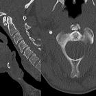
Bogenspalt im
Dornfortsatz BWK 1 bei einer jungen Frau. Computertomographie axial und in der Volumen-Rekonstruktion.


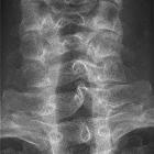
Längerstreckige
sakrale Dysraphie im Röntgenbild und in der Computertomografie (Volumen Rendering): Einen nicht komplett geschlossenen knöchernen dorsalen Wirbelkörperbogen sieht man im Bereich des Sacrums nicht selten, hier längerstreckig über das ganze Sacrum. Nebenbefundlich Phlebolithen im Becken beidseits.


Unfused
spinous process • Unfused spinous processes of C6 and C7 - Ganzer Fall bei Radiopaedia

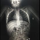
Bogenspalt am
BWK 2 im Röntgenbild. Zufallsbefund ohne Klinik. Beachte auch die Form des Dornfortsatzes von HWK 7.

Lateral
sacral meningocele presenting as a gluteal mass: a case report. X-ray, anteroposterior view of the lumbosacral spine showing L5 and S1 spina bifida.


Unfused
spinous process • Unfused spinous process of C7 - Ganzer Fall bei Radiopaedia

Diastematomyelia
• Diastematomyelia - type II - Ganzer Fall bei Radiopaedia

Spalt am
hinteren Atlasbogen bei einem über 70-jährigen. Korrespondierend findet sich im vorderen Bogen eine mediane Sklerosierung. Aber kein echter Split-Atlas.
Spina bifida
• Spina bifida - Ganzer Fall bei Radiopaedia

Bogenspalt am
BWK 2 im Röntgenbild. Zufallsbefund ohne Klinik. Beachte auch die Form des Dornfortsatzes von HWK 7.

Spina bifida
• Spina bifida - Ganzer Fall bei Radiopaedia

Spina bifida
• Spina bifida - Ganzer Fall bei Radiopaedia

Spina bifida
• Spina bifida with lipomyelomeningocoele - Ganzer Fall bei Radiopaedia

Spina bifida
• Spina bifida occulta - Ganzer Fall bei Radiopaedia

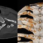
Bogenspalt im
HWK 6 bei einem 68-jährigen. Computertomographie links axial (von unten gesehen) und rechts als Volumen-Rendering (schräg von oben hinten).

Spina bifida
• Hip dislocation and spina bifida - Ganzer Fall bei Radiopaedia

Spina bifida
• Charcot arthropathy of the knee - Ganzer Fall bei Radiopaedia
Unfused spinous process, which is really failure of fusion of the neural arch, is a relatively common anatomical variant and is part of the spectrum of spina bifida occulta.
This should be differentiated from accessory ossicles of the spinous process, which appear after non-fusion of the secondary ossification center (or centers in bifid spinous processes). These appear as a well-corticated fragment adjacent to the tip of the spinous process with a vertical or near-vertical lucency.
The importance lies in patients with potentially traumatic spinal injuries, in which it is important to identify this variant and avoid potential pitfall of diagnosing a fracture. A well-defined cortical margin is a helpful clue.
Differential diagnosis
- spinous process accessory ossicles
- spinous process fracture
Siehe auch:
- Atlasbogenspalt
- Varianten des hinteren Atlasbogens
- Arnold-Chiari-Malformation Typ 2
- Schmetterlingswirbel
- Pätau-Syndrom
- Hüftdysplasie
- angeborene Wirbelanomalien
- lemon sign
- Klumpfuß
- Meningozele
- Trisomie 18
- fetal ventriculomegaly
- Myelomeningozele
- Roberts-Syndrom
- Aicardi-Goutières-Syndrom
- Varianten des vorderen Atlasbogens
- neuroenterische Zyste
- Triploidie
- Fetales Valproat-Syndrom
- neurogene Blase
- Talus verticalis
- Neuralrohrdefekt
- koronare Wirbelspalten
- Bananenzeichen (Kleinhirn)
- rachischisis
- Dysrhaphie
- kongenitaler Hydrozephalus
- zervikaler Bogenspalt
- increased maternal alpha feto protein (MSAFP)
- Spina bifida Sakrum
und weiter:


 Assoziationen und Differentialdiagnosen zu Spina bifida: