dysembryoplastic neuroepithelial tumor


Dysembryoplastic neuroepithelial tumors (DNET) are benign (WHO Grade I) slow growing glioneuronal tumors arising from either cortical or deep grey matter. The vast majority are centered in cortical grey matter, arise from secondary germinal layers, and are frequently associated with cortical dysplasia (in up to 80% of cases). They characteristically cause intractable partial seizures (see temporal lobe epilepsy).
Epidemiology
Typically these tumors are diagnosed in children or young adults, as a result of the investigation of seizures, which have usually had childhood onset. Only a slight male predilection is present . An association with Noonan syndrome has been proposed .
Clinical presentation
Patients with DNETs typically present with longstanding treatment-resistant partial seizures (in 90% of cases the first seizure occurred before the age of 20 ) without associated or progressive neurological deficit .
Pathology
Location
DNETs are most often located in the temporal lobe although all parts of the CNS containing grey matter are potential locations.
- temporal lobe: over ~65% of cases
- frontal lobe: ~20% of cases
- caudate nucleus
- cerebellum: present more commonly with ataxia rather than seizures
- pons
Macroscopic appearance
Macroscopically, DNETs are visible on the surface of the brain, sometimes with an exophytic component. When sectioned they demonstrate heterogeneous, often gelatinous, cut surface with nodules of firmer tissue .
Microscopic appearance
DNETs are a mixed glioneuronal neoplasm with a multinodular architecture and a heterogeneous cellular composition. The "specific glioneuronal element (SGNE)" is characteristic, and refers to columnar bundles of axons surrounded by oligodendrocyte-like cells which are oriented at right angles to the overlying cortical surface. Between these columns are "floating neurons" as well as stellate astrocytes .
Three histological forms are recognized :
- SGNE only
- SGNE, with...
- glial nodules and a multinodular architecture
Focal cortical dysplasia is commonly seen in association with DNETs, and unless a component can be identified clearly separate from tumor cells, then it does not warrant a concurrent separate diagnosis. If, however, such a separate component is present, then it represents Blumcke classification IIIb focal cortical dysplasia) .
Immunophenotype
The stellate astrocytes within the SGNE are positive for GFAP .
The oligodendrocyte-like cells are typically S100 and OLIG2 positive, and may also express NOGO-A and myelin-oligodendrocyte glycoprotein .
The floating neurons are positive for NeuN .
Importantly, DNETs are negative for IDH mutations, TP53 mutations, and do not demonstrate 1p19q co-deletion . These features are helpful in distinguishing DNETs from low-grade astrocytomas (usually IDH mutated) and oligodendrogliomas (IDH mutated and 1p19q co-deleted).
Radiographic features
DNETs are typically predominantly cortical and well-circumscribed tumors.
CT
DNETs appear as low-density masses, usually with no or minimal enhancement. When cortical, as is usually the case, they may scallop/remodel the inner table of the skull vault but without erosion. In some cases, the cranial fossa can be minimally enlarged at times.
Calcification is visible in ~30% (more common histologically) and is typically visualized in the deepest parts of the tumor, particularly adjacent to enhancing or hemorrhagic areas .
MRI
Typically seen as a cortical lesion with hardly any surrounding vasogenic edema.
- T1
- generally hypointense compared with adjacent brain
- T1 C+ (Gd)
- may show enhancement in ~20-30% of cases
- enhancement may be heterogeneous or a mural nodule
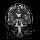
- T2
- generally high signal
- high signal 'bubbly appearance'
- FLAIR
- mixed-signal intensity with bright rim sign
- partial suppression of some of the "bubbles"
- FLAIR is helpful in identifying the small peripheral lesions with similar intensity to CSF
- T2*
- calcification relatively frequent
- hemosiderin staining uncommon as bleeding into DNETs is only occasional
- DWI
- no restricted diffusion
- MR spectroscopy
- non-specific although lactate may be present
Treatment and prognosis
They demonstrate essentially no growth over time, although very gradual increase in size has been described. Only one case of malignant transformation has been reported .
Prognosis is excellent, however, due to the difficulty in managing seizure medically, patients usually undergo resection and even in cases of incomplete resection, seizures frequently cease.
Differential diagnosis
Main differential diagnosis is that of other cortical tumors, with helpful distinguishing features including :
- ganglioglioma
- contrast enhancement more common
- calcification in ~50% of cases
- no 'bubbly' appearance
- pleomorphic xanthoastrocytoma (PXA)
- contrast enhancement prominent
- dural tail sign often seen
- diffuse low-grade astrocytoma
- IDH mutated
- lacking histological specific glioneuronal element (SGNE)
- oligodendroglioma
- IDH mutated and 1p19q co-deleted
- lacking histological specific glioneuronal element (SGNE)
- desmoplastic infantile astrocytomas and ganglioglioma
- young children
- dural involvement prominent
- large often multiple lesions
Importantly the 'bubbly' appearance can be seen also in multinodular and vacuolating neuronal tumors (MVNT) which are however in the juxtacortical white matter, rather than in the cortex .
The differential diagnosis also depends on the location of the tumor.
Temporal lobe consider:
- tumors (in order of decreasing frequency)
- cysts
- other
- herpes simplex encephalitis: usually some bilateral changes, and different presentation
- limbic encephalitis: usually some bilateral changes, and different presentation
- mesial temporal sclerosis (MTS)
See also: temporal lobe tumors
If cortical elsewhere consider:
- low-grade astrocytoma
- ganglioglioma
- pleomorphic xanthoastrocytoma (PXA)
- oligoastrocytoma/oligodendroglioma
- Taylor dysplasia
Siehe auch:
- Pilozytisches Astrozytom
- Oligodendrogliom
- Gangliogliom
- Astrozytom
- Pleomorphes Xanthoastrozytom
- Hippocampussklerose
- Heterotopie der grauen Substanz
- Herpesenzephalitis
- Limbische Enzephalitis
- multinodulärer und vakuolisierender neuronaler Tumor
- diffuses Astrozytom
- Taylor type cortical dysplasia
- Oligoastrozytome
- Tumoren des Temporallappens
- benigne Raumforderung des cerebralen Kortex
und weiter:


 Assoziationen und Differentialdiagnosen zu Dysembryoplastischer neuroepithelialer Tumor:


