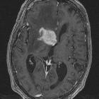
dural tail sign


The dural tail sign occurs as a result of thickening and enhancement of the dura and is most often seen adjacent to a meningioma.
Initially, the sign was felt to be pathognomonic of meningiomas, however as radiologist's experience grew, it has become increasingly noted to be present in many other conditions, although without the same regularity:
- meningioma (by far the most common, seen in 52-78% of cases)
- pleomorphic xanthoastrocytoma
- glioblastoma
- chloroma
- dural plasmacytoma
- primary CNS lymphoma
- sarcoidosis
- vestibular schwannoma
- cerebral metastases
- syphilitic gumma
- medulloblastoma
- desmoplastic infantile astrocytoma and ganglioglioma
- pituitary adenoma
A useful mnemonic can be found here.
Pathology
Initially, the dural tail was thought to result from direct invasion of the dura by the tumor, however, subsequent studies demonstrated it to be predominantly a reactive process due to vascular congestion and edema. Having said that, the literature is still divided and a wide range of prevalence of tumor invasion of the dural tail has been reported (0-100%), with generally higher prevalences in WHO II (atypical) meningiomas . This is further complicated by the presence of tumor cells in apparently normal dura adjacent to tumors .
Treatment and prognosis
Whether or not the dural tail should be resected and if so how much surrounding dura should be included in the resection (or gamma knife field) continues to be debated .
History and etymology
It was first described in 1989 by Wilms et al. as thickening of the dura surrounding meningiomas .
Siehe auch:
- Meningeom
- Sarkoidose
- Hirnmetastase
- Glioblastoma multiforme
- Medulloblastom
- primäres ZNS-Lymphom
- Pleomorphes Xanthoastrozytom
- Vestibularisschwannom
- Myelosarkom
- infantiles desmoplastisches Gangliogliom
- dural plasmacytoma
- syphylitische Gumma
und weiter:


 Assoziationen und Differentialdiagnosen zu Dural-Tail-Zeichen: