mitral stenosis


Mitral valve stenosis is a valvulopathy that describes narrowing of the opening of the mitral valve between the left ventricle and the left atrium.
Epidemiology
Mitral stenosis is seen more commonly in women and in countries, generally developing nations, where rheumatic fever is common .
Clinical presentation
Patients with mitral stenosis characteristically present with progressive dyspnea that is precipitated by sudden changes in heart rate, volume status or cardiac output (e.g. physical exertion, fever, emotional stress, anemia, atrial fibrillation, pregnancy, etc.) . Clinical examination classically reveals a malar flush ('mitral facies') due to cutaneous vasoconstriction, and a mid-diastolic murmur that is heard on praecordial auscultation . Auscultation may also reveal an opening snap and a loud first heart sound .
Patients may also manifest symptoms from complications of mitral stenosis, such as hemoptysis from pulmonary venous hypertension, or Ortner syndrome from mass-effect of the large left atrium, or those of heart failure .
ECG
- left atrial enlargement
- may precipitate atrial fibrillation
- right ventricular hypertrophy
- concomitant left atrial enlargement without left ventricular hypertrophy is especially suggestive
Pathology
Mitral stenosis is usually acquired via rheumatic heart disease, where there is chronic inflammation of the mitral valve leaflets (mitral valvulitis) . This leads to progressive and diffuse fibrous thickening of the valve leaflets, and development of valvular calcifications . Eventually, the mitral commisures fuse and the chordae tendinae fuse . This culminates in significant immobilization and narrowing of the mitral valve, giving it a characteristic 'fish mouth' appearance . Many patients will also have concurrent mitral regurgitation due to the valve being unable to sufficiently close .
The characteristic hemodynamic feature of mitral stenosis is an increased left atrial pressure . This increase in pressure is required as a compensatory mechanism for the stenosis, in order to maintain normal cardiac output . However, this compensation results in left atrial enlargement and an increase in pulmonary venous pressure . An increase in pulmonary venous pressure eventually leads to the development of pulmonary arterial hypertension, explaining why dyspnea and hemoptysis are such prominent and important symptoms in mitral stenosis .
However, this adaptive mechanism eventually fails because as the left atrial pressure continues to increase as the stenosis worsens, the amount of time needed to fill the left ventricle with blood also increases . This can be compounded by atrial fibrillation, a complication of left atrial enlargement, which results in the loss of the 'atrial kick' at the end of diastole and an even greater left atrial pressure being needed .
This becomes particularly problematic if there is an increase in heart rate (i.e. aforementioned precipitants) because the diastolic period shortens more than the systolic period . This means there is even less time to fill the left ventricle, often resulting in a sudden drop in cardiac output and development of acute pulmonary edema .
Etiology
In addition to being a sequela of rheumatic fever, which is the most common cause world-wide, there are numerous other causes :
- mitral annular calcification with leaflet involvement
- an age-related cause
- congenital mitral stenosis
- infective endocarditis
- cor triatriatum
- connective tissue disorders
- radiation-induced heart disease
- left atrial myxoma (generally not considered 'true' mitral stenosis)
- ball valve thrombus (generally not considered 'true' mitral stenosis)
Radiographic features
Plain radiograph
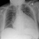
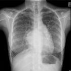
Typical chest radiographic features include :
- left atrial enlargement
- convexity or straightening of the left atrial appendage just below the main pulmonary artery (along left heart border)
- double density sign: the right side of the enlarged left atrium pushes into the adjacent lung and creates an addition contour superimposed over the right heart
- elevation of the left main bronchus and splaying of the carina
- walking man sign on lateral projections
- upper zone venous enlargement due to pulmonary venous hypertension
- pulmonary edema
- diffuse alveolar hemorrhage
- secondary pulmonary hemosiderosis, often difficult to appreciate on a plain radiograph
- pulmonary ossification, a late sign
If the underlying etiology is mitral annular calcification, then this may also be appreciated on plain film.
Ultrasound: echocardiography
Echocardiography is useful for assessing the mitral valve area, jet velocity, pressure gradients, and the left ventricle . Various parameters are used in order to determine severity, such as :
- mild
- valve area >1.5 cm
- mean gradient <5 mmHg
- pulmonary artery pressure <30 mmHg
- moderate
- valve area 1.0-1.5 cm
- mean gradient 5-10 mmHg
- pulmonary artery pressure 30-50 mmHg
- severe
- valve area <1.0 cm
- mean gradient >10 mmHg
- pulmonary artery pressure >50 mmHg
Additionally, the mitral valve anatomy can be assessed . One widely adopted echocardiographic scoring system for this is one that Wilkins et al. developed.In this system, four features of the mitral valve are identified, as follows :
- valve leaflet mobility
- valve leaflet thickening
- valve leaflet calcification
- subvalvular thickening
CT
CT confirms, with greater detail, findings on plain radiograph such as left atrial enlargement, features of heart failure, and secondary pulmonary hemosiderosis .
Dynamic CT imaging
Rheumatic mitral stenosis may have distinctive morphological features on dynamic imaging . Restricted opening of the thickened valve from commissural fusion (especially with rheumatic valve disease), valve leaflet calcification, or both, results in a 'fish mouth' appearance on short-axis images . Bowing of a thickened and fibrotic anterior leaflet during diastole may result in a 'hockey-stick' appearance which is best seen on two- or four-chamber images .
On the other hand, non-rheumatic causes of mitral stenosis usually produce nonspecific imaging features such as valve thickening or leaflet fixation .
MRI
Cardiac MRI (CMR) is able to provide the most detailed structural and dynamic assessment of the mitral valve and left-sided cardiac chambers .
Cine MRI
Observable features include :
- mitral leaflet thickening
- reduced diastolic opening
- abnormal valve motion toward the left ventricular outflow tract
VEC-MRI
Velocity-encoded cine-magnetic resonance imaging (VEC-MRI) is a relatively new method for quantitation of blood flow with the potential to measure high-velocity jets across stenotic valves .
Treatment and prognosis
The decision to treat mitral stenosis is based on the severity and presence of complications. Management involves a combination of lifestyle and pharmacotherapy measures (a similar armamentarium to that used in heart failure and atrial fibrillation), and mitral valve surgery (e.g. mitral valvotomy or mitral valve replacement) . Additionally, patients with rheumatic mitral stenosis may need penicillin prophylaxis to prevent recurrence .
Further details in regards to management is beyond the scope of this article.
Complications
- congestive heart failure
- pulmonary hypertension
- mass-effect from left atrial enlargement (e.g. Ortner syndrome, dysphagia megalatriensis)
- pulmonary embolus (often recurrent)
- pulmonary infections (e.g. pneumonia, bronchitis)
- atrial fibrillation
- sudden cardiac death from other arrhythmias
- thrmoboembolic ischemic stroke, including from calcified cerebral embolism if the cause is mitral annular calcification
- mitral regurgitation
- tricuspid regurgitation
See also
- valvular heart disease
- mitral valve disease
- general mitral valve pathologies:
- mitral valve stenosis
- mitral valve regurgitation
- specific mitral valve pathologies:
- congenital mitral valve stenosis
- cor triatriatum
- mitral annular calcification
- mitral valve atresia
- mitral valve prolapse
- parachute mitral valve
Siehe auch:
- mitral annular calcification
- rheumatic heart disease
- Mitralklappenverkalkungen
- vergößerter linker Vorhof
und weiter:

 Assoziationen und Differentialdiagnosen zu Mitralklappenstenose:


