PAN


Polyarteritis nodosa (PAN) is a systemic inflammatory necrotizing vasculitis that involves small to medium-sized arteries (larger than arterioles).
Epidemiology
PAN is more common in males and typically presents around the 5 to 7decades. 20-30% of patients are hepatitis B antigen positive.
Associations
Clinical presentation
Patients can present with systemic and focal symptoms.
Non-specific systemic signs and symptoms are almost always present and include fever, malaise and weight loss.
Localized symptoms relate to ischemia and infarction of affected tissues and organs. The most commonly involved vessels are the renal arteries , with visceral involvement also considered relatively common. The pulmonary circulation is typically spared, although bronchial arteries may occasionally be involved.
Frequent sites of involvement are :
- renal: 80-90%, tends to be the prominent site and major cause of death
- cardiac: ~70%
- gastrointestinal tract: 50-70%
- hepatic: 50-60%
- spleen: 45%
- pancreas: 25-35%
- CNS complications: 20-45%
Pathology
Initially, there is transmural and necrotizing inflammation of medium-sized arteries, mostly involving part of the circumference which causes weakening of the wall leading to microaneurysm formation and subsequent focal rupture. There is a predilection for branch points. Fibrinoid necrosis of vessels promotes thrombosis of vessels followed by infarction of the tissue supplied. Fibrous thickening and mononuclear infiltration occur at a later stage. Different stages of inflammation can occur in the same vessel at different points.
Markers
- pANCA: it has been claimed that pANCA levels may correlate with disease activity, although the utility of this marker has been debated
Radiographic features
CT
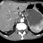
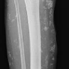
Routine contrast-enhanced CT may be entirely normal or may demonstrate focal regions of infarction or hemorrhage in affected organs.
Angiography (DSA / CT angiography)
Direct catheter angiography is far more sensitive to changes within small vessels, although a good quality CTA can also demonstrate changes. Findings include:
- multiple microaneurysms
- characteristic but not pathognomonic
- typically 2-3 mm in size but can be up to 1 cm
- in the kidneys, the microaneurysms typically involve the interlobar and arcuate arteries
- hemorrhage may be present due to focal rupture
- occlusion may be present
Treatment and prognosis
Polyarteritis nodosa is usually fatal if untreated, often as a result of progressive renal failure or gastrointestinal complications. Prompt treatment with corticosteroids and cyclophosphamide may result in remission, and a remission/cure can be achieved in 90% of patients .
History and etymology
It was initially described by Kussmaul and Maier in 1866.
Differential diagnosis
- consider other vasculitides such as
- microscopic polyangiitis: has a much more established association with ANCA and tends to affect smaller arterioles, capillaries and venules
- rheumatoid vasculitis
- systemic lupus erythematosus (SLE)
- Churg-Strauss syndrome
Siehe auch:
- Churg-Strauss-Syndrom
- Vaskulitis
- Aortitis
- systemischer Lupus Erythematodes
- Mikroskopische Polyangiitis
- renale Mikroaneurysmen
- renale Mikroaneurysmen bei Polyarteriits nodosa
- Polyarteriitis nodosa Angiographie
und weiter:
- Aneurysma spurium
- Ileitis terminalis
- inflammatorischer Pseudotumor der Orbita
- Nierenarterienstenose
- Aneurysma Koronararterien
- causes of pulmonary arterial hypertension
- posttraumatisches Aneurysma spurium
- Wunderlich-Syndrom
- chronic bilateral airspace opacification
- renovaskuläre arterielle Hypertonie
- hypertrophe Pachymeningitis
- eosinophile Granulomatose mit Polyangiitis
- Gefäßbeteiligung bei Polyarteriitis nodosa
- aphthoid ulceration
- sekundär sklerosierende Cholangitis
- segmentale arterielle Mediolyse (SAM)
- Arteriitis
- Vaskulitis im Gastrointestinaltrakt
- Vaskulitis der Gallenblase
- chronische Periaortitis

 Assoziationen und Differentialdiagnosen zu Polyarteriitis nodosa:


