Rule of 4 of the brainstem

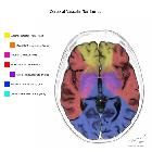
The rule of 4 of the brainstem elegantly simplifies and explains the anatomy of the brainstem and the basis for various brainstem stroke syndromes. This article summarizes the original four rules , associated important clinical deficits, important exceptions, and provides two examples of their clinical utility.
Rules
Rule 1
There are 4 cranial nerves from above the pons (including 2 from the midbrain), 4 from the pons, and 4 from the medulla oblongata:
- from above the pons
- CN I (olfactory nerve): not from the midbrain, essentially a peripheral outpost of the central nervous system
- CN II (optic nerve): not from the midbrain, essentially a peripheral outpost of the central nervous system
- CN III (oculomotor nerve): from the midbrain, involvement leads classic 'down and out’ appearance with the eye resting in abduction, slight depression and intorsion due to paralysis of adduction, elevation and depression, the pupil is often not involved (see oculomotor nerve palsy)
- CN IV (trochlear nerve): from the midbrain, involvement leads to the eye unable to look down when the eye is looking inferomedially (e.g. walking downstairs, reading, etc.), there is also head tilt away from the affected eye
- from the pons
- CN V (trigeminal nerve): involvement leads to ipsilateral loss of pain and temperature on the face, as well as also the ipsilateral weakness of muscles of mastication
- CN VI (abducens nerve): involvement leads to ipsilateral weakness of abduction of the eye (see abducens nerve palsy)
- CN VII (facial nerve): involvement leads to ipsilateral facial weakness, loss of taste in the anterior two-thirds of the tongue, and hyperacusis
- CN VIII (vestibulocochlear nerve): involvement leads to ipsilateral deafness
- importantly, the vestibular nuclei are located in the lateral medulla, involvement of which leads to vertigo, vomiting and nystagmus , although intractable vomiting can also occur due to damage to the area postrema in the dorsomedial aspect of the medulla
- from the medulla oblongata
- CN IX (glossopharyngeal nerve): involvement leads to ipsilateral loss of sensation of the tonsils, pharynx, middle ear, and posterior one-third of the tongue, there may be weakness in swallowing
- CN X (vagus nerve): involvement leads to ipsilateral palatal weakness and associated deviation of the uvula, loss of the gag reflex may also be present
- CN XI (accessory nerve): involvement leads to ipsilateral weakness in head rotation and elevation of shoulder (sternocleidomastoid and trapezius muscles respectively)
- CN XII (hypoglossal nerve): involvement leads to ipsilateral weakness of the tongue (deviates to side of lesion)
Rule 2
The 4 cranial nerve motor nuclei that are in the mid-line (actually paramedian) are those that divide equally into 12 except CN I and II:
- mid-line (actually paramedian) 4 motor nuclei: those of CN III, IV, VI, and XII
- lateral motor nuclei: those of CN V, VII, IX, X, and XI
- no motor nuclei: CN I, II, VIII
Rule 3
There are 4 ‘mid-line’ (i.e. medial, but actually paramedian) structures beginning with ‘m’:
- importantly, in the midbrain, the medial lemniscus is located more lateral than in the pons and comes together with the spinothalamic pathway
- importantly, does not extend down to the medulla oblongata in the same clinical relevance
Rule 4
There are 4 ‘side’ (i.e. lateral) structures beginning with ‘s’:
- an important caveat to this is in the midbrain, where involvement of the red nucleus (a medial midbrain structure) can also cause ataxia (albeit contralateral) and choreoathetosis (compare medial midbrain stroke syndromes Benedikt syndrome or Claude syndrome that involve the red nucleus with Weber syndrome that does not)
- importantly, does not extend up into the midbrain, and thus lesions of the midbrain spinothalamic pathway will cause total contralateral loss of pain, temperature, vibration, and proprioception (due to the medial lemniscus fibers also being more lateral in the midbrain)
Examples
Although Gates' rules simplify the anatomy of the brainstem, they are still very useful when approaching brainstem stroke syndromes . Furthermore, these rules can also be applied to clinical features developed as a result other brainstem lesions, such as demyelination, external brainstem compressions (e.g. cerebellopontine angle masses), and brainstem tumors .
Importantly, not all of the features as suggested by the rule of 4 of the brainstem may be present in every brainstem stroke syndrome, as evident in many midbrain stroke syndromes in particular (as seen in above exceptions), but often only some are needed to localize the lesion . Furthermore, if both medial and lateral brainstem signs are detected clinically, then lesions such as a basilar artery occlusion (for pontine and midbrain syndromes) or hemimedullary syndrome from proximal vertebral artery occlusion should be suspected .
Example 1
Working backward for the sake of demonstration, if a patient is considered, using Gates' rule of 4 of the brainstem, with a known left-sided medial medullary syndrome (Dejerine syndrome).
Thus, a patient with left-sided Dejerine syndrome is likely to present with right-sided limb weakness, right-sided loss of limb vibration and proprioception, and left-sided tongue weakness and atrophy.
Example 2
Working to make a diagnosis as in real-life, if a patient is considered, using Gates' rule of 4 of the brainstem, presenting with right-sided deafness, ataxia, Horner syndrome, paralysis of the face, loss of pain and temperature from the face, and left-sided loss of pain and temperature in the limbs.
Thus, this constellation of clinical features is consistent with right-sided lateral pontine syndrome (Marie-Foix syndrome).
History and etymology
The original rule of 4 of the brainstem was devised and published by Peter Gates, an Australian neurologist, in 2003 in order to simply explain the anatomy of the brainstem and basis of brainstem stroke syndromes to the non-neurologist and medical student .

