tuberculomas of the brain and spinal cord


Intracranial tuberculous granulomas, also known as CNS tuberculomas, are common in endemic areas and may occur either in isolation or along with tuberculous meningitis.
Epidemiology
The epidemiology of patients with tuberculomas is the same as that of other CNS manifestations of tuberculosis (TB) (see CNS tuberculosis).
Pathology
A tuberculoma is distinct from a tuberculous abscess in that it demonstrates evidence of granulomatous reaction and caseous necrosis histologically. In contrast, abscesses do not display a granulomatous reaction and their centers are filled with pus . Not all tuberculomas, however, have a solid granulomatous core and some may undergo liquefaction . Tuberculous organisms may not necessarily be identified in tuberculomas, whereas they are necessary to make the diagnosis of tuberculous abscess .
Radiographic features
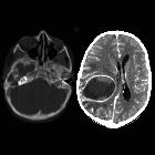
CT
On CT, tuberculomas may appear as a round or lobulated nodule with moderate to marked edema. Either solid or ring enhancement is typical post-contrast. A central focus of calcification with a ring of peripheral enhancement (the "target sign") is described but is not specific to tuberculosis . When calcification is present (the minority of cases) it tends to be larger than that calcification seen in neurocysticercosis.
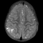
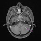
MRI
MRI is the modality of choice in assessing potential tuberculomas which have fairly solid caseous necrosis centrally on the background of granulomatous reaction. In some instances, however, liquefactive necrosis centrally can occur, and the imaging appearances are then essentially indistinguishable from a tuberculous abscess, which in turn is similar to pyogenic cerebral abscesses .
- T1
- isointense to grey matter
- may have a central region of hyperintensity representing caseation
- T2
- isointense to grey matter
- may have a central region of hypointensity representing gliosis and abundant monocyte infiltration
- lesions are surrounded by vasogenic edema
- T1 C+ (Gd)
- usually appears as ring-enhancement
- may appear as a conglomerate enhancing mass
- DWI
- typically central low signal (i.e. no restricted diffusion) but if liquid necrosis is present centrally may be high signal
- MR spectroscopy
- decrease in NAA/Cr
- slight decrease in NAA/Cho
- lipid-lactate peaks are usually elevated (86%)
Differential diagnosis
The differential of tuberculomas is primarily the differential of ring-enhancing lesions, and includes:
- other infection
- neurosarcoidosis
- cerebral metastases
- CNS lymphoma
Central isointensity or hypointensity compared to grey matter seen centrally on T2 is helpful, as it is not seen in most other causes .
Siehe auch:
- Meningeosis neoplastica
- Meningeom
- Hirnmetastase
- meningeale Kontrastmittelaufnahme
- Neurosarkoidose
- Neurozystizerkose
- Tuberkulose des ZNS
- Neurotoxoplasmose
- Erdheim-Chester-Erkrankung
- zerebrale Kryptokokkose
- ZNS Lymphom
- zerebrale Läsionen mit ringförmiger Kontrastmittelanreicherung
- Kleinhirnabszess
- Läsionen mit ringförmiger Kontrastmittelanreicherung
- bakterieller Hirnabszess
- tuberkulöse Meningitis
- cerebellar tuberculoma
- multifocal spinal tuberculosis
- zerebrale Miliartuberkulose
- ZNS-Manifestationen bei Langerhans-Zell-Histiozytose
- miliary cerebral tuberculosis
- intrakranieller tuberkulöser Abszess
- cerebellar tuberculomas


 Assoziationen und Differentialdiagnosen zu Tuberkulom des ZNS: