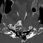
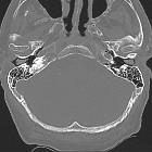
tympanic cavity


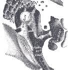
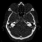
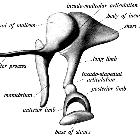


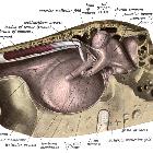
The middle ear or middle ear cavity, also known as tympanic cavity or tympanum (plural: tympanums/tympana), is an air-filled chamber in the petrous part of the temporal bone. It is separated from the external ear by the tympanic membrane, and from the inner ear by the medial wall of the tympanic cavity. It contains the three auditory ossicles whose purpose is to transmit and amplify sound vibrations from the tympanic membrane to the oval window of the lateral wall of the inner ear.
Gross anatomy
The tympanic cavity is subdivided into several parts defined in relation to the planes of the tympanic membrane. Some authors define three compartments :
- mesotympanum, directly medial to the membrane
- epitympanum (attic, epitympanic recess), superior to the membrane
- hypotympanum, inferior to the membrane
In addition to these compartments, some authors define two more compartments :
- protympanum, anterior to the membrane
- retrotympanum, posterior to the membrane
The middle ear is shaped like a narrow box with concave sides. It has six "walls":
Contents
Bones
Middle ear ossicles consist of three small bones (the malleus, incus and stapes), which form a mobile chain across the tympanic cavity from the tympanic membrane to the oval window.
Muscles
There are two muscles, one attached to the malleus and one attached to the stapes, which act to damp down over-vibration from low-pitched sound waves.
- tensor tympani muscle (inserts into the handle of the malleus)
- stapedius muscle (inserts into the neck of the stapes)
Nerves
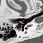
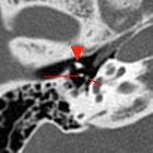
The chorda tympani , a branch of nervus intermedius, leaves the facial nerve in the facial canal and enters the tympanic cavity through the posterior wall, lateral to the pyramid, lying just underneath the mucous membrane. It runs over the pars flaccida of the tympanic membrane, and the neck of the malleus. It leaves at the anterior margin of the tympanic notch.
Specific reconstructions as Stenvers view or double-oblique sagittal view can be useful to assess the involvement of the facial canal in tumors affecting the tympanic cavity, as well as in pre-surgical assessment .
Arterial supply
- anterior tympanic artery from the maxillary artery
- stylomastoid artery from the posterior auricular or occipital arteries
- numerous small vessels from the external carotid artery
Venous drainage
- drainage to the pterygoid venous plexus and the superior petrosal sinus
Lymphatic drainage
Lymphatic drainage is to the parotid, retropharyngeal and upper group of deep cervical nodes.
Innervation
This is by the tympanic branch of the glossopharyngeal nerve (Jacobson nerve), which forms the tympanic plexus by combining with sympathetic fibers from the internal carotid nerve. Branches from the plexus supply sensory and vasomotor fibers to the mucous membrane of the tympanic cavity, as well as to the tympanic membrane and external auditory meatus.
The middle and external ear are also supplied by branches of the trigeminal, facial, glossopharyngeal and vagus nerves which results in referred pain in the ear from other areas supplied by these nerves, e.g. the teeth, posterior part of the tongue, pharynx and larynx.
The tympanic plexus gives off the lesser petrosal nerve, which provides parasympathetic innervation to the parotid gland via the otic ganglion.
History and etymology
Tympanum is derived from τύμπανον (tumpanon) the Ancient Greek word for a drum.
Related pathology
- middle ear granulation tissue
- otomastoiditis
- middle ear effusion
- hemotympanum
- tympanic membrane retraction
- eustachian tube dysfunction
- middle ear tumors
- cholesterol granuloma
- acquired cholesteatoma
Siehe auch:
- Cholesterolgranulom der Felsenbeinspitze
- Tuba auditiva Eustachii
- Felsenbein
- Ossikel
- Trommelfell
- erworbenes Cholesteatom
- Mastoiditis
- Gehörknöchelchen
- eustachian tube dysfunction
- Innenohr
- middle ear effusion
- tympanic membrane retraction
- middle ear granulation tissue
- Processus cochleariformis
- external ear
- Tumoren des Mittelohres
und weiter:
- Bulbus-venae-jugularis-Hochstand
- Cholesteatom
- Nervus facialis
- aberrante Arteria carotis interna
- cochleariform process
- Paragangliom
- inner ear
- Cholesteatom des äußeren Gehörgangs
- oval window
- Chorda tympani
- Nervus glossopharyngeus
- Labyrinthitis
- Fenestra cochleae rotunda
- Paragangliom des Nervus vagus
- dehiszenter Bulbus jugularis
- Schwannom des Nervus facialis
- Paragangliome von Kopf und Hals
- Promontorium (Mittelohr)
- Osteom des Mittelohrs

 Assoziationen und Differentialdiagnosen zu Cavum tympani:


