external carotid artery


The external carotid artery (ECA) is one of the two terminal branches of the common carotid artery. The other terminal branch is the internal carotid (ICA), which is somewhat larger than the ECA.
Summary
- origin: bifurcation of the common carotid artery
- course: under the submandibular gland and into the parotid gland
- branches:
- supply: neck, face and base of skull
- termination: division into (internal) maxillary artery and superficial temporal artery
Gross anatomy
Origin and course
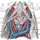
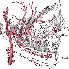
The ECA begins at the level of the upper border of the thyroid cartilage (at the level of the fourth cervical vertebra). It takes a slightly curved course upwards and anteriorly before inclining backwards to the space behind the neck of the mandible. Along its course, it rapidly diminishes in size and as it does so, gives off various branches (see below). As it enters the parotid gland, it gives rise to its terminal branches, the superficial temporal and maxillary arteries.
Branches
The branches of the external carotid artery can be subdivided into groups:
- arising from the carotid triangle
- posterior auricular artery
- terminal branches
Memorable mnemonics for these branches include:
- Some Anatomists Like Freaking Out Poor Medical Students
- Some American Ladies Found Our Pyramids Most Satisfactory
Relations
- anteriorly (i.e. ECA is crossed by these structures)
- upper root of ansa cervicalis
- hypoglossal nerve (CN XII)
- posterior belly of digastric muscle
- stylohyoid muscle and ligament
- facial nerve (CN VII) (within the parotid gland)
- retromandibular vein (within the parotid gland)
- passing between ECA and ICA
- pharyngeal branch of vagus nerve (CN X)
- glossopharyngeal nerve (CN IX)
- stylopharyngeus muscle
- styloglossus muscle
- posteriorly (i.e. ECA lies on these structures)
- pharyngeal wall
- superior laryngeal branch of vagus nerve (CN X)
- deep lobe of the parotid gland
Variant anatomy
- variations in origin arise from the anomalous bifurcation of the common carotid artery - bifurcations may commonly be seen at the level of the cricoid cartilage (C5 level) or at the hyoid bone (C2 level)
- variant branching patterns
- linguofacial trunk (incidence ~20%): common origin lingual and facial arteries
- thyrolingual trunk (incidence ~2.5%): common origin superior thyroid and lingual arteries
- thyrolinguofacial trunk (incidence ~2.5%): common origin superior thyroid, lingual and facial arteries
- common occipito-auricular trunk (incidence ~12.5%): common origin occipital and posterior auricular arteries
Siehe auch:
- Arteria carotis communis
- Arteria pharyngea ascendens
- Glandula parotidea
- Arteria maxillaris
- Glandula submandibularis
- Arteria lingualis
- Arteria facialis
- Arteria occipitalis
- posterior auricular artery
- Mandibula
- Arteria thyroidea superior
- mnemonics
- bifurcation
- internal carotid (ICA)
und weiter:
- Arteria carotis interna
- Glomus caroticum Tumor
- aberrante Arteria carotis interna
- parotid space
- persistierende karotidobasiläre Anastomosen
- carotid body
- hot nose sign
- carotid bifurcation
- Hirntod
- congenital absence of the internal carotid artery
- Arteria carotis
- Glomus caroticum
- persistent proatalantal intersegmental artery
- Embryologie Arteria basilaris
- Äste der Arteria maxillaris
- moyamoya staging (Susuki)
- Carotis-Sinus-cavernosus-Fistel
- Rami caroticotympanici

 Assoziationen und Differentialdiagnosen zu Arteria carotis externa:


