Myelomeningozele

Newborn with
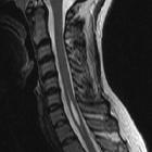
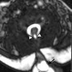
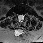
a myelomeningocele. Sagittal T1 MRI without contrast of the brain (left) shows a small posterior fossa with downward cerebellar tonsil herniation and a small fourth ventricle. There is kinking of the spinal cord at the cervico-medullary junction. Sagittal (above right) and axial (below right) T2 MRI without contrast of the spine shows a low-lying conus medullaris with the spinal cord nerve roots terminating in a posteriorly located cerebrospinal fluid filled sac which is not covered by skin at the level of the L5-S1 vertebral bodies.The diagnosis was Chiari II malformation with a myelomeningocele.

Myelomeningocele
• Chiari II malformation - Ganzer Fall bei Radiopaedia
Myelomeningocele
• Myelomeningocoele in an adult - Ganzer Fall bei Radiopaedia

Myelomeningocele
• Myelomeningocele - Ganzer Fall bei Radiopaedia

Myelomeningocele
• Myelomeningocele - Ganzer Fall bei Radiopaedia

Myelomeningocele
• Chiari II malformation with spinal meningomyelocele - Ganzer Fall bei Radiopaedia
Myelomeningocele
• Chiari II malformation - Ganzer Fall bei Radiopaedia

Preschooler
who had their myelomeningocele extending from L4-S5 repaired in utero. 3D reconstruction of CT without contrast of the lumbar spine shows lack of fusion of the posterior elements of the L4-S5 vertebral bodies.The diagnosis was myelomeningocele.

Banana sign
(cerebellum) • Myelomeningocoele - Ganzer Fall bei Radiopaedia

Tethered cord
syndrome • Chiari type II malformation - on ultrasound - Ganzer Fall bei Radiopaedia

Chiari II
malformation • Upper thoracic spinal dysraphism - Ganzer Fall bei Radiopaedia

Chiari II
malformation • Chiari II malformation - Ganzer Fall bei Radiopaedia
Chiari II
malformation • Chiari II malformation - Ganzer Fall bei Radiopaedia
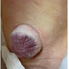
Myelomeningocele, also known as spina bifida cystica, is a complex congenital spinal anomaly that results in spinal cord malformation (myelodysplasia).
Epidemiology
It is one of the commonest congenital CNS anomalies and thought to occur in approximately 1:500 of live births . There may be a slight female predilection.
Clinical presentation
Patients present with lower limb paralysis and sensory loss, bladder and bowel dysfunction as well as cognitive impairment.
Pathology
Results from failure of fusion of neural tube dorsally during embryogenesis.
There is a localized defect of the closure of caudal neuropore with persistence of neural placode (open spinal cord).
Location
- lumbosacral: ~45%
- thoracolumbar: ~30%
- lumbar: ~20%
- cervical: ~2%
Risk factors
- in utero folate deficiency
- in utero teratogen exposure
Markers
Associations
- aneuploidy anomalies
- Chiari II malformation
- diastematomyelia
- syringomyelia
- arachnoid cysts
- hydrocephalus
- tethering of spinal cord
Radiographic features
Antenatal ultrasound
- may show evidence of an open neural tube defect with splayed or divergent posterior elements
Siehe auch:
- Arachnoidalzyste
- Spina bifida
- Hydrocephalus
- Arnold-Chiari-Malformation Typ 2
- Pätau-Syndrom
- Meningozele
- Trisomie 18
- Syringomyelie
- Diastematomyelie
- Lipomeningomyelozele
- Dysrhaphie
- Myelozystozele
- raised maternal alpha feto-protein (AFP)
und weiter:
- Chiari-Malformation Typ 1
- Syrinx
- Tethered cord
- cyllosoma
- lipomyelomeningocele
- neuroradiologisches Curriculum
- Steißbeinteratom
- Myeloschisis
- Chiari-Malformation
- Kloakenekstrophie
- erweiterter Spinalkanal
- kongenitale Kniegelenksluxation
- Läsionen des Sakrums
- Erweiterung des interpedunculären Abstands
- myelomeningocele with dorsal dermal sinus

 Assoziationen und Differentialdiagnosen zu Myelomeningozele:


