CDH


Congenital diaphragmatic herniation (CDH) accounts for a small proportion of all diaphragmatic herniae. However, it is one of the most common non-cardiac fetal intrathoracic anomalies.
Epidemiology
Congenital diaphragmatic hernias are seen in 1 of every 2000-4000 live births. 84% are left-sided, 13% are right-sided and 2% bilateral .
Clinical presentation
Most congenital diaphragmatic hernias are detected either soon after birth or on antenatal ultrasound. Mortality is predominantly due to the development of pulmonary hypoplasia, which is thought to be due to mass effect on the developing lung. Such neonates are hypoxic and have persistent fetal circulation due to pulmonary hypoplasia and pulmonary hypertension.
Pathology
Diaphragmatic development is usually complete by ~9 week of gestation. Congenital diaphragmatic hernias result from failure of fusion of one of the pleuroperitoneal canals at about 8 weeks gestation. They may contain stomach, intestines, liver or spleen.
Classification
Congenital diaphragmatic herniation can be classified into two basic types on location:
- most common fetal congenital diaphragmatic hernia
- commoner on the left: 75-90%
- posterolateral
- large and associated with poorer outcome
- presents earlier
- mnemonic: BBBBB
- less common
- anterior
- presents later
Associations
While a CDH can occur as an isolated condition, associated anomalies are relatively common and include:
- pulmonary hypoplasia: also a complication
- bronchopulmonary sequestration
- aneuploidy: can be present in up to 50% of cases
- Fryns syndrome
- Cornelia de Lange syndrome
- congenital cardiac anomalies
- neural tube defects
Radiographic features
Plain radiograph
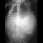
- indistinct diaphragm with opacification of part of or all the hemithorax (typically left sided)
- scaphoid abdomen
- deviation of lines
- endotracheal tube
- nasogastric tube
- umbilical arterial and venous catheters
Antenatal ultrasound
Indirect sonographic findings that should prompt a search for CDH include :
- polyhydramnios
- cardiomediastinal shift +/- abnormal cardiac axis
- inability to demonstrate the normal stomach bubble
The study should be performed in the true transverse plane. Sonographic diagnosis of CDH can be made from the following findings :
- absent bowel loops in the abdomen
- intrathoracic herniation of the liver; noted in up to 85% of cases and is associated with a worse prognosis
- peristaltic bowel movements in the chest
- herniation into the chest may occur intermittently
- abdominal circumference is reduced (due to herniation of organs)
- left-sided CDH
- stomach and small bowel (echo-free) at the same transverse level as the heart on four-chamber view: this makes left sided hernias comparatively easier to detect on ultrasound (as opposed to herniation of echogenic liver on the right side)
- stomach and small bowel superior to the inferior margin of the scapula
- leftward displacement of the gallbladder
- right-sided CDH
- color Doppler study
- leftward bowing of the umbilical segment of the portal vein
- portal branches to the lateral segment of the left hepatic lobe coursing towards or above the diaphragm
- gallbladder present above the diaphragm
- echogenic space between the left heart border and stomach representing the left hepatic lobe
- color Doppler study
Although classically considered a cystic echogenic lung mass, there are reports of CDH appearing initially as a solid echogenic lung mass that evolves in appearance with advancing gestation .
The observed-to-expected lung-to-head ratio (O/E LHR) may be calculated and correlates with the degree of pulmonary hypoplasia. Studies suggest that the degree of lung hypoplasia can be used to predict survival rates and the numbers from the Antenatal-CDH-Registry group that apply to isolated left-sided CDH and liver herniation are shown below :
- O/E LHR < 15% (extreme pulmonary hypoplasia): virtually no chance of survival
- O/E LHR 15-25% (severe pulmonary hypoplasia): predicted survival ≈ 15%
- O/E LHR 26-45% (moderate pulmonary hypoplasia): predicted survival 30-75%
- O/E LHR > 45% (mild pulmonary hypoplasia): very likely to survive
MRI
Fetal MRI may be helpful in further assessing the hernia and any associated pulmonary hypoplasia.
Sequences typically performed for assessment of CDH include :
- T2 weighted three-plane single shot fast spin echo (SSFSE)
- fluid filled stomach and small bowel appear hyperintense
- T2 weighted balanced steady state free precession (bSSFP)
- flowing blood appears hyperintense: portal vessels may be seen extending toward or above the diaphragm
- T1 weighted fast field echo (FFE)
- liver appears moderately hyperintense
- T2 weighted half-fourier acquisition single-shot turbo spin echo (HASTE)
- lungs appear hyperintense (composed primarily of water) while heart, mediastinum and liver appear hypointense
Lung-head ratio: can be assessed on both ultrasound or MRI; MRI measured LHR has been found to have a slightly higher prognostic accuracy than with ultrasound .
MRI allows the measurement of fetal lung volumes which provide an estimate of the severity of pulmonary hypoplasia. The total fetal lung volume (TFLV) can be calculated from a contiguous T2-weighted HASTE sequence and an observed-to-expected TFLV (O/E TFLV) derived. It has been found to predict well both mortality and morbidity, including the need for ECMO and the development of bronchopulmonary dysplasia .
Complications
- development of pulmonary hypoplasia
- development of pulmonary hypertension
- severity may be predicted by the modified McGoon index
Treatment and prognosis
Fetuses with an antenatal diagnosis of CDH should be delivered in a tertiary referral center with access to neonatal intensive care and pediatric surgical facilities.
Large CDH have a poor prognosis, due to pulmonary hypoplasia and perinatal mortality may be as high as 80%. Clearly, successful management is dependent on specialist pediatric facilities, with the ability to offer surgery, ECMO etc...
Signs suggesting a poor prognosis include:
- large hernia size
- early gestational age at diagnosis
- intra-thoracic liver
- small contralateral lung
- the presence of associated abnormalities
- bilateral CDH
- unfavorable lung: head ratio
A composite prognostic index (CDH-CPI) comprising 10 prenatal parameters has been developed and was found to have a stronger correlation with survival and need for ECMO than any one parameter individually .
Some centers perform in utero surgery in selected cases, including novel methods such as clamping the fetal trachea to allow expansion of the fetal lungs with fluid and consequently push the herniation back into the abdomen.
Differential diagnosis
General imaging differential considerations include:
- congenital pulmonary airway malformation
- hybrid lesion
- pulmonary sequestration (can also be an association)
Siehe auch:
- Lungensequester
- kongenitale pulmonale Atemwegsmalformation (CPAM)
- Zwerchfellhernie
- Cornelia-de-Lange-Syndrom
- Fryns-Syndrom
- Lungensequester extralobulär
- Cantrell’sche Pentalogie
- hybrid lesion: CCAM - pulmonary sequestration
- traumatische Zwerchfellruptur
- Zwerchfelldefekte
- Zwerchfellagenesie
- kongenitale Hernien
- Fused adrenal gland
und weiter:
- upside-down-Magen
- Hydrops fetalis
- Zwerchfell
- nuchal translucency
- Polyhydramnion
- fetal pleural effusion
- interstitielles Lungenemphysem
- organo-axial gastric volvulus
- neonatal chest radiograph in the exam setting
- Magenvolvulus
- mesenteroaxialer Magenvolvulus
- PIE
- cytic lung lesions - paediatric
- differential diagnosis of an absent fetal stomach on ultrasound
- Herzektopie
- Acute Respiratory Distress Syndrome neonatal


 Assoziationen und Differentialdiagnosen zu kongenitale Zwerchfellhernie: