pylorospasm


Hypertrophic pyloric stenosis (HPS) refers to the idiopathic thickening of gastric pyloric musculature which then results in progressive gastric outlet obstruction.
Epidemiology
Pyloric stenosis is relatively common, with an incidence of approximately 2-5 per 1,000 births, and has a male predilection (M: F ~4:1). It is more commonly seen in Caucasians and is less common in India and among black and other Asian populations.
Risk factors
- being firstborn
- maternal history of pyloric stenosis
Clinical presentation
While symptoms may start as early as 3 weeks, it typically clinically manifests between 6 to 12 weeks of age. Clinical presentation is typical with non-bilious projectile vomiting. The hypertrophied pylorus can be palpated as an olive-sized mass in the right upper quadrant. A succussion splash may be audible, and although common, is only relevant if heard hours after the last meal . Due to the loss of hydrochloric acid in the gastric contents from persistent vomiting, patients are at risk of electrolyte imbalance, specifically the characteristic hypochloremic metabolic alkalosis.
Pathology
Pyloric stenosis is the result of both hyperplasia and hypertrophy of the pyloric circular muscle fibers. The pathogenesis of this is not understood. There are four main theories :
- immunohistochemical abnormalities
- genetic abnormalities
- infectious cause
- hyperacidity theory
Associations
- Turner syndrome
- tracheo-esophageal fistula
- esophageal atresia
- trisomy 18
Radiographic features
Plain radiograph
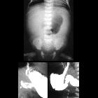
Abdominal x-ray findings are non-specific but may show a distended stomach with minimal distal intestinal bowel gas.
Fluoroscopy
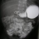
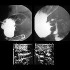
An upper gastrointestinal series (barium meal) excludes other, more serious causes of pathology, but the findings of an upper gastrointestinal series infer, rather than directly visualize, the hypertrophied muscle. On upper gastrointestinal fluoroscopy:
- delayed gastric emptying
- peristaltic waves (caterpillar sign)
- elongated pylorus with a narrow lumen (string sign) which may appear duplicated due to puckering of the mucosa (double-track sign)
- the pylorus indents the contrast-filled antrum (shoulder sign) and (tit sign) or base of the duodenal bulb (mushroom sign)
- the entrance to the pylorus may be beak-shaped (beak sign)
Ultrasound
Ultrasound is the modality of choice in the right clinical setting because of its advantages over a barium meal are that it directly visualizes the pyloric muscle and does not use ionizing radiation. Unfortunately, it is incapable of excluding other diagnoses such as midgut volvulus. Easy ultrasound technique is to find gallbladder then turn the probe obliquely sagittal to the body in an attempt to find pylorus longitudinally .
The hypertrophied muscle is hypoechoic, and the central mucosa is hyperechoic. Diagnostic measurements include (mnemonic "number pi"):
- pyloric muscle thickness, i.e. diameter of a single muscular wall (hypoechoic component) on a transverse image: >3 mm (most accurate )
- length, i.e. longitudinal measurement: >15-17 mm
- pyloric volume: >1.5 cm
- pyloric transverse diameter: >13 mm
With the patient's right side down the pylorus should be watched and should not be seen to open.
Described sonographic signs include:
Treatment and prognosis
Initial medical management is essential with rehydration and correction of electrolyte imbalances. This should be completed prior to surgical intervention.
Treatment is surgical with a pyloromyotomy in which the pyloric muscle is divided down to the submucosa. This can be performed both open and laparoscopically. The operation is curative and has very low morbidity . Recurrence is rare and usually due to an incomplete pyloromyotomy .
Differential diagnosis
There is usually little differential when imaging findings are appropriate. Of course, clinically it is important to consider other causes of vomiting in infancy.
A degree of pylorospasm is common in infancy and is responsible for some delay in gastric emptying. The pylorus, however, appears sonographically normal. In cases where the doubts persist, fluid gastric distention can be performed to "open" a tapered pylorus.
Gastro-esophageal reflux which represents the cause of vomiting in two-thirds of infants referred to radiology .
Other causes of proximal gastrointestinal obstruction can be considered :
See also
Siehe auch:
- Pancreas anulare
- Dünndarmvolvulus
- Duodenalstenose
- duodenales Web
- Antral nipple sign (pyloric stenosis)
- antral nipple sign
- cervix sign of pyloric stenosis
- shoulder sign of pyloric stenosis
- target sign of pyloric stenosis
- mushroom sign of pyloric stenosis
- Roviralta-Syndrom
- gastrointestinal string sign
- double track sign of pyloric stenosis
- beak sign in pyloric stenosis
- Magenausgangsstenose bei Kindern
und weiter:


 Assoziationen und Differentialdiagnosen zu hypertrophe Pylorusstenose: