anterosuperior mediastinal masses


Getting a film with an anterior mediastinal mass in the exam is one of the many exam set-pieces that can be prepared for.
The film goes up and after a couple of seconds pause, you need to start talking:
CXR
There is a left sided mediastinal mass that makes obtuse angles with the mediastinal contour. The hilar vessels can be seen through the mass - this is the hilum overlay sign and means this is not in the middle mediastinum. The paravertebral line can also be seen, placing this mass in the anterior mediastinum.
The differential includes lymphoma, thyroid malignancy, thymoma and teratoma and the age of the patient and accompanying clinical symptoms and signs would help order the list.
I would notify the referring clinician urgently and suggest a CT scan to assist diagnosis and assess the extent of disease.
CT
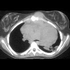
Selected axial and sagittal slices from a contrast enhanced CT chest.
This confirms that the mass is in the anterior mediastinum. There are fatty and calcific components suggesting a diagnosis of teratoma. A CT-guided biopsy could be performed to confirm the diagnosis.
Notes
- when the CT goes up - don't go back to the beginning. Use the phrase "this confirms..."
- where there is fat and calcification in an anterior mediastinal mass, think teratoma
- in lymphoma, Hodgkin's disease is more common in younger patients with lymphadenopathy isolated to the chest
See also
Siehe auch:
- Mediastinum
- Thymus
- Teratom
- Lymphom
- Perikard
- Chorionkarzinom
- Schilddrüse
- Keimzelltumor
- mediastinales Teratom
- Morbus Hodgkin
- thorakales Aortenaneurysma
- Normale Herzkonfiguration im Röntgen-Thorax
- anterosuperior mediastinal mass (mnemonic)
- vorderes Mediastinum
- Neoplasien der Schilddrüse
- Tumoren des Thymus
- mediastinales Seminom
- IgG4-assoziierte Erkrankung des Mediastinums
- hilum overlay sign
- chest radiograph in the exam setting
und weiter:
- Pancoast tumour
- Thymom
- Thymuskarzinom
- Morbus Castleman
- mediastinal lymphoma
- mediastinale Raumforderungen
- Thymuslipom
- enlargement of the cardiac silhouette
- retrosternal airspace
- invasives Thymom
- mediastinale Keimzelltumoren
- large-cell lymphoma of the mediastium
- obliteration of the retrosternal airspace
- oberes Mediastinum
- thymic epithelial tumours
- Raumforderungen oberes Mediastinum
- adult chest radiograph set-pieces
- Teratom des Thymus
- Metastasen im Thymus
- Karzinoid des Thymus
- mediastinal malignant germinoma
- lymphofollikuläre Thymushyperplasie
- hydatid cyst of the mediastinum
- Chondrosarkom des Sternums
- primäres Lymphom des Thymus

 Assoziationen und Differentialdiagnosen zu Tumoren des vorderen oberen Mediastinums:


