cervical carcinoma


Carcinoma of the cervix is a malignancy arising from the cervix. It is the third most common gynecologic malignancy (after endometrial and ovarian).
Epidemiology
It typically presents in younger women with an average age of onset at around 45 years.
Risk factors
- human papillomavirus (HPV) 16 and 18 infections: for most types except for clear cell carcinoma of the cervix and mesonephric carcinoma of the cervix
- multiple sexual partners or a male partner with multiple previous or current sexual partners
- young age at first intercourse
- high parity
- immunosuppression
- certain HLA subtypes
- oral contraceptives
- nicotine/smoking (except for cervical adenocarcinoma )
Clinical presentation
Presenting symptoms include:
- vaginal bleeding
- vaginal discharge
- subclinical: an abnormal cervical cancer screening test
Pathology
Invasive cervical carcinoma is thought to arise from the transformation of cervical intraepithelial neoplasia (CIN).
Histological types
The main histological types are:
- squamous cell carcinoma of the cervix: accounts for the vast majority (80-90%) of cases and is associated with exposure to human papillomavirus (HPV)
- adenocarcinoma of the cervix: rarer (5-20%) and can have several subtypes which include
- clear cell carcinoma of the cervix
- endometroid carcinoma of the cervix: ~7% of adenocarcinomas
- mucinous carcinoma of the cervix
- adenoma malignum: ~3% of adenocarcinomas
- serous carcinoma of the cervix
- mesonephric carcinoma of the cervix: ~3% of adenocarcinomas
- neuroendocrine tumors of the cervix
- small cell carcinoma of the cervix: rare (0.5-6%)
- adenosquamous cell carcinoma of the cervix: rare
For a detailed overview, refer to:
Location
Cervical squamous cell carcinoma arises from the squamocolumnar junction while adenocarcinomas arise from the endocervix. The squamocolumnar junction is situated on the ectocervix in younger patients though regresses into the endocervical canal with age. Hence cervical tumors tend to be exophytic in younger patients and endophytic with advancing age.
Radiographic features
General features
In order to be radiographically visible, tumors must be at least stage Ib or above (see staging). MRI is the imaging modality of choice to depict the primary tumor and assess the local extent. Distant metastatic disease is best assessed with CT or PET, where available.
Although the FIGO staging system is clinically based, the revised 2009 FIGO staging encourages imaging as an adjunct to clinical staging. MRI can stratify patients to the optimum treatment group of primary surgery or combined chemotherapy and radiotherapy. Tumors stage IIa and below are treated with surgery.
Ultrasound
- hypoechoic, heterogeneous mass involving the cervix
- may show increased vascularity on color Doppler
- although cervical cancer is staged clinically, ultrasound can be a useful adjunct by showing
- size (<4 cm or >4 cm)
- parametrial invasion
- tumor invasion into the vagina
- tumor invasion into adjacent organs
- hydronephrosis: implies stage IIIB tumor.
CT
CT, in general, is not very useful in the assessment of the primary tumor, but it can be useful in assessing the advanced disease. It is performed primarily to assess adenopathy, but also has roles in defining advanced disease, monitoring distant metastasis, planning the placement of radiation ports, and guiding percutaneous biopsy.
On CT, the primary tumor can be hypoenhancing or isoenhancing to normal cervical stroma (~50% ).
PET-CT
PET-CT in conjunction with pelvic MRI is often used as an imaging strategy in helping stage cervical carcinoma.
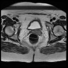
MRI
A dedicated MRI protocol is often useful for optimal imaging assessment.
The normal low signal cervical stroma provides intrinsic contrast for the high signal cervical tumor.
- T1: usually isointense compared with pelvic muscles
- T2
- hyperintense relative to the low signal of the cervical stroma
- hyperintensity is thought to be present regardless of histological subtype
- T1 C+ (Gd)
- contrast is not routinely used, though it may be helpful to demonstrate small tumors considered for trachelectomy
- on contrast-enhanced T1-weighted images, tumor presents as a high signal relative to the low signal of the cervical stroma
For further information, see the article: MRI reporting guidelines for cervical cancer.
Staging
The FIGO staging system is the most commonly adopted. See: cervical cancer staging
Treatment and prognosis
Prognosis is affected by many factors which include:
- tumor stage
- the volume of the primary mass
- histologic grade
Five-year survival rates vary between 92% for stage I disease and 17% for stage IV disease .
One of the keys roles of the radiologist is to help determine staging, as this may lead to appropriate management pathway either with surgery or chemo-radiotherapy. At the time of writing stage IIa vs. IIb is considered as an important separator in deciding whether a case is operable or not.
Differential diagnosis
For a mass involving the cervix consider:
- cervical polyp
- cervical leiomyoma
- invasion of the cervix from
- cervical lymphoma
- adenoma malignum: often considered a subtype of mucinous carcinoma of the cervix
- metastases to the cervix
- cervical ectopic pregnancy: consider with women of childbearing age with a high βHCG
Practical points
- MRI T2WI to assess parametrial invasion (stage 2b) is crucial to determine if the patient is candidate for surgery or not
Siehe auch:
- cervical ectopic pregnancy
- Vaginalkarzinom
- Adenoma malignum der Zervix uteri
- maligne Uterustumoren
- Zervixkarzinom Staging
- MRI protocol for assessment of cervical carcinoma
- Leiomyom der Zervix uteri
- Metastasen in der Cervix uteri
- Ultrastaging
- Lymphom der Zervix uteri
- stage 0 cervical cancer
und weiter:
- cancer
- Endometriumkarzinom
- FIGO-Klassifikation
- AIDS defining illness
- sonographic values in obstetrics and gynaecology
- Vulvakarzinom
- AIDS defining malignancies
- clear cell carcinoma of the cervix
- gemischt osteolytisch osteoblastische Knochenmetastasen
- trachelectomy
- Plattenepithelkarzinom der Zervix uteri
- gynäkologisch radiologisches Curriculum
- mesonephric carcinoma of the cervix
- squamo-columnar junction of cervix
- small cell carcinoma of the cervix
- Brachytherapie
- papillary serous carcinoma of the cervix
- Zervikale intraepitheliale Neoplasie
- transposed ovaries
- adeno squamous cell carcinoma of the cervix
- pelvic MRI protocol cervical carcinoma
- Lungenmetastasen bei Zervixkarzinom
- WHO Klassifikation der Tumoren der Cervix uteri
- Epitheloidsarkom der Cervix uteri
- Adenokarzinom der Cervix uteri
- Seeds Brachytherapie
- endometroides Adenokarzinom der Cervix uteri


 Assoziationen und Differentialdiagnosen zu Zervixkarzinom: