fibroids


Uterine leiomyomas also referred to as uterine fibroids, are benign tumors of myometrial origin and are the most common solid benign uterine neoplasms. Commonly an incidental finding on imaging, they rarely cause a diagnostic dilemma. There are various medical, surgical, and interventional treatment options.
Epidemiology
They occur in ~25% of women of reproductive age and are particularly common in the African population.
Fibroids are responsive to hormones (e.g. stimulated by estrogens). Being rare in prepubertal females, they commonly accelerate in growth during pregnancy and involute with menopause .
Clinical presentation
They are often asymptomatic and discovered incidentally. Signs and symptoms associated with fibroids include:
- abnormal vaginal bleeding
- pain
- infertility
- palpable masses
Pathology
Leiomyomas are benign monoclonal tumors predominantly composed of smooth muscle cells with variable amounts of fibrous connective tissue. They are commonly multiple (~85% ), and range significantly in size.
Fibroids may have a number of locations within or external to the uterus:
- intra-uterine
- intramural leiomyoma: most common
- subserosal leiomyoma
- submucosal leiomyoma: least common (10-15%)
- extra-uterine
- diffuse uterine leiomyomatosis
Subserosal fibroids may be pedunculated and predominantly extra-uterine, simulating an adnexal mass. Any fibroid may undergo atrophy, internal hemorrhage, fibrosis, and calcification.
They can also undergo several types of degeneration:
- hyaline degeneration: focal or generalized hyalinisation: this is the most common type of degeneration (can occur in ~60% of cases)
- cystic degeneration: ~5%
- myxoid degeneration: generally considered uncommon although reported as high as 50% by some authors
- red/carneous degeneration: due to hemorrhagic infarction, which can occur particularly during pregnancy, and may present with acute abdominal pain
Based on histology
Radiographic features
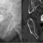
Plain radiograph
Popcorn calcification within the pelvis may suggest the diagnosis.
Ultrasound
Ultrasound is used to diagnose the presence and monitor the growth of fibroids:
- uncomplicated leiomyomas are usually hypoechoic, but can be isoechoic, or even hyperechoic compared to normal myometrium
- calcification is seen as echogenic foci with shadowing
- cystic areas of necrosis or degeneration may be seen
- Venetian blind artefact may be seen but edge shadowing +/- dense posterior shadowing from calcification is also typically seen
CT
- fibroids are usually seen as soft tissue density lesions and may exhibit coarse peripheral or central calcification
- they may distort the usually smooth uterine contour
- enhancement pattern is variable
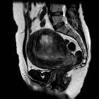
MRI
MRI is not generally required for diagnosis, except for complex or problem-solving cases. It is, however, the most accurate modality for detecting, localizing, and characterizing fibroids. Size, location, and signal intensity should be noted.
Signal characteristics are variable and include :
- T1
- non-degenerated fibroids and calcification appear as low to intermediate signal intensity compared with the normal myometrium
- characteristic high signal intensity on T1 weighted images/an irregular, T1 hyperintense rim around a centrally located myoma suggests red degeneration, which is caused by venous thrombosis
- T2
- non-degenerated fibroids and calcification appear as low signal intensity
- as they are usually hypervascular, flow voids are often observed around them
- fibroids that have undergone cystic degeneration/necrosis can have a variable appearance, usually appearing high signal on T2 sequences.
- hyaline degeneration is demonstrated as low T2 signal intensity
- cystic degeneration, which is an advanced stage of intratumoral edema, also shows high signal intensity on T2 weighted images and does not enhance
- T1 C+ (Gd)
- variable enhancement is seen with contrast administration
- the marked high signal intensity with gradual enhancement (albeit mild) suggests myxoid degeneration
MRI is of significant value in the symptomatic patient when surgery and uterine salvage therapy is considered. It is also of great value in differentiating a pedunculated fibroid from an adnexal mass.
Treatment and prognosis
Common treatment options include:
- myomectomy
- focal endometrial curettage
- hormone administration
- hysterectomy
- uterine artery embolization (UAE)
Complications
- rarely invasion of adjacent venous channels leading to intravenous leiomyomatosis
- rarely (0.1-0.5%), they undergo malignant degeneration into leiomyosarcomas
- in extremely rare instances, lesions capable of metastasizing without malignant transformation: benign metastasizing leiomyoma
- fibroids may torse, leading to acute pelvic pain
- pregnancy may cause fibroid growth by 30%
Differential diagnosis
General imaging differential considerations include:
- uterine leiomyosarcoma
- malignant transformation into leiomyosarcoma is rare.
- unfortunately, no imaging modality can reliably differentiate a benign leiomyoma from the rare leiomyosarcoma
- uterine smooth muscle tumors of uncertain malignant potential: rare
- uterine lipoleiomyoma: greater fat content (sometimes considered a variant of a leiomyoma)
- focal myometrial contraction (Braxton Hicks contraction): especially if seen during pregnancy
- focal adenomyosis: less well-defined; color Doppler demonstrates tortuous vessels through the abnormality; no calcifications
In occasional situations, it may be difficult to differentiate between uterine leiomyomas and:
- ovarian or adnexal masses (especially if the leiomyoma is pedunculated)
- endometrial carcinoma
Siehe auch:
- Blasenstein
- Adenomyose
- Lipoleiomyom des Uterus
- Uterine smooth muscle tumors of uncertain malignant potential
- subseröses Uterusmyom
- intra-mural uterine leiomyoma
- Uterus myomatosus
- Leiomyom der Vagina
- Uterusmyom-Embolisation
- Braxton Hicks contraction
- Leiomyosarkom des Uterus
- nekrotisch verändertes Uterusmyom
- submuköses Leiomyofibrom des Uterus
und weiter:
- Uterus
- endometrial polyp
- verkalktes Uterusmyom
- Hysterosalpingographie
- endometrial hyperplasia
- popkornartige Verkalkungen
- Endometriumkarzinom
- ovarian artery
- carneous degeneration of a leiomyoma
- diffuse uterine leiomyomatosis
- adenomyotic cyst
- differential diagnosis of an enlarged uterus
- uterine perforation
- Braxton Hicks contractions
- fetal akinesia / hypokinesia sequence
- hereditary leiomyomatosis and renal cell carcinoma
- abdominelle und pelvine Verkalkungen
- large for dates uterus
- Vorzeitige Plazentalösung
- Leiomyom der Harnblase
- Adenomyom
- Chorioangiom der Plazenta
- gynäkologisch radiologisches Curriculum
- pelvine extrauterine Leiomyome
- Leiomyom der Zervix uteri
- Leiomyom mit Verkalkungen
- Leiomyofibrom
- Embolisation Arteria uterina
- Zervixpolypen
- eccentric gestational sac
- Arteria uterina
- calcified uterine fibroma
- uterine artery embolisation : MRI assessment
- Leiomyomatosis peritonealis disseminata
- zellreiches Leiomyom des Uterus
- intraabdominelle Verkalkungen
- Leiomyom des Uterus mit zystischer Degeneration
- Rhabdomyosarkom des Uterus
- HIFU
- Prolaps eines submukösen Leiomyofibroms des Uterus
- Leiomyom des Uterus mit hyaliner Degeneration


 Assoziationen und Differentialdiagnosen zu Leiomyofibrom Uterus: