neurofibromas


Neurofibromas are benign peripheral nerve sheath tumors usually solitary and sporadic, however, there is a strong association with neurofibromatosis type 1 (NF1). These tumors present as a well-defined hypodense mass with minimal or no contrast enhancement on CT. On MRI, they usually are T1 hypointense and T2 hyperintense with heterogeneous contrast enhancement.
Neurofibromas are generally divided into three subtypes :
- localized neurofibroma
- localized cutaneous neurofibroma
- localized intraneural neurofibroma
- diffuse cutaneous neurofibroma (superficial)
- plexiform neurofibroma (deep)
Diffuse cutaneous neurofibromas and plexiform neurofibromas are discussed separately, and localized cutaneous neurofibromas are generally not a radiological diagnosis. As such, the remainder of this article is a general discussion focusing on the most common localized intraneural neurofibromas which are by far the most common form of neurofibroma, representing 90% of these lesions .
Epidemiology
Peak presentation is between 20 and 30 years of age with no sex predilection.
Associations
The majority of localized intraneural neurofibromas are solitary and sporadic and not associated with neurofibromatosis type 1. However, when multiple neurofibromas are present (or plexiform neurofibromas) then the diagnosis of NF1 is almost assured.
Clinical presentation
The clinical presentation of localized intraneural neurofibromas is non-specific and the result of either mass effect on surrounding lesions (or palpable lump) or neurogenic dysfunction.
Pathology
Localized intraneural neurofibromas are benign neoplasms composed of Schwann cells and fibroblasts, containing a rich network of collagen fibers. They are mostly indolent tumors that rarely only undergo malignant transformation into a malignant peripheral nerve sheath tumor (MPNST); this is only really seen in large neurofibromas, and even then it only occurs in 5-10% of tumors .
Neurofibromas are therefore considered WHO grade I tumors under the current (2016) WHO classification of CNS tumors .
Macroscopic appearance
Unlike schwannomas, neurofibromas are not encapsulated and infiltrate between the nerve fascicles. They primarily affect superficial cutaneous nerves, however occasionally affect larger deep-seated nerves.
Radiographic features
General imaging features of neurofibromas:
CT
- well-defined hypodense mass
- minimal or no contrast enhancement
MRI
- T1: hypointense
- T2
- hyperintense
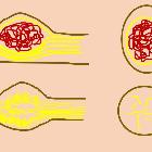
- target sign
- a hyperintense rim and central area of a low signal may be seen
- this is thought to be due to a dense central area of collagenous stroma
- although this sign is highly suggestive of neurofibroma, it is occasionally also seen in schwannomas and malignant peripheral nerve sheath tumors
- fascicular sign
- T1 C+ (Gd): heterogeneous enhancement
Treatment and prognosis
- lesions not associated with NF1
- localized and diffuse lesions may be treated surgically
- however, as neurofibromas infiltrate between nerve fascicles, they are unable to be separated from the parent nerve and complete excision requires the sacrifice of the nerve
- deep-seated lesions are therefore often managed conservatively
- local recurrence after excision is uncommon and malignant transformation is rare
- lesions associated with NF1
- due to the multiplicity of lesions, unless debilitating symptoms are present, treatment of patients with NF1 is often non-surgical
- plexiform neurofibromas are particularly difficult to resect, often leading to incomplete resection.
- recurrence after resection is frequent
- plexiform neurofibromas demonstrate a significant potential for malignant transformation
Siehe auch:
- Neurofibromatose Typ 1
- Schwannom
- spinal neurofibroma
- plexiformes Neurofibrom
- Unterscheidung Schwannom Neurofibrom
- localized neurofibroma
und weiter:
- Myelonkompression
- laterale Halszyste
- Epidermoidzyste
- genitourinary curriculum
- Skoliose
- carotid space
- WHO-Klassifikation der Tumoren des zentralen Nervensystems
- spinale Schwannome
- neuroradiologisches Curriculum
- maligner peripherer Nervenscheidentumor (MPNST)
- Steißbeinteratom
- Tumoren des hinteren Mediastinums
- posterior mediastinal masses
- broad ligament leiomyoma
- Tumoren der Thoraxwand
- oberflächliche Weichteilläsionen der Extremitäten
- Zyste oder Fistel des zweiten Kiemenbogens
- benigner peripherer Nervenscheidentumor
- paraspinal ganglioneuroma
- Fasciitis nodularis
- posterior mediastinal mass in the exam
- Weichteiltumoren der Extremitäten
- Infantile Myofibromatose
- Zyste oder Fistel des vierten Kiemenbogens
- paraspinal mass
- Vergrößerung der Glandula parotis
- Läsionen der Mandibula
- sporadisches Neurofibrom
- localised neurofibroma
- diffuse neurofibroma
- neurogenic tumours
- Nervenscheidentumor
- Fibrom
- Schwannom des Nervus facialis
- zervikale Mittellinienraumforderungen
- neurofibroma of the ischial nerve
- intraparotid neurofibroma
- neurofibroma of the ankle
- phrenic nerve neurofibroma
- Faszikel-Zeichen
- pelvines Neurofibrom


 Assoziationen und Differentialdiagnosen zu Neurofibrom: