differential for an anterosuperior mediastinal mass

Thymuskarzinom
in der Computertomographie mit Kontrastmittel. Der maligne Charakter wird durch die ummauernde Ausdehnung und unscharfe (infiltrierende!?) Begrenzung deutlich. 1 : Das Karzinom 2 : V. cava superior 3 : Truncus brachiocephalicus 4 : Linke A. subclavia und A. carotis communis 5 : Aortenbogen 6 : Sternum

Malignes
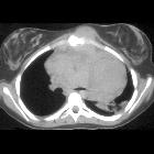
Thymom / Thymuskarzinom in der Computertomographie (axial). Die Histologie erbrachte ein primäres Karzinom des Thymus vom Plattenepithelial-Typ, dem häufigsten Typ des Thymuskarzinoms. Das CT zeigt eine große, nicht scharf begrenzte Formation im vorderem Mediastinum mit Verdrängung der Lunge und Zeichen einer möglichen Infiltration in Nachbarstrukturen. Inhomogene Kontrastierung. Zysten oder Verkalkungen fanden sich in diesem Fall nicht.

Malignes
Thymom / Thymuskarzinom in der Computertomographie (sagittal). Die Histologie erbrachte ein primäres Karzinom des Thymus vom Plattenepithelial-Typ, dem häufigsten Typ des Thymuskarzinoms. Das CT zeigt eine große, nicht scharf begrenzte Formation im vorderem Mediastinum mit Verdrängung der Lunge und Zeichen einer möglichen Infiltration in Nachbarstrukturen. Inhomogene Kontrastierung. Zysten oder Verkalkungen fanden sich in diesem Fall nicht.

Mikronoduläres
Thymom mit lymphoidem Stroma. Hier Computertomographie axial.

Anterior
mediastinal mass consistent with primary mediastinal large B cell lymphoma. Axial chest CT shows an enhancing left anterior mediastinal mass compressing the aortic arch.

Mediastinal
liposarcoma of thymic origin. Axial CECT Thorax shows heterogeneously enhancing soft tissue density mass in the anterior mediastinum with areas of fat (*) displacing the trachea (T) slightly to the right.

Mediastinal
liposarcoma of thymic origin. Coronal CECT showing the mass lesion and pericardial effusion (e). Fat components (*). Mild displacement of trachea (T) to the right is noted.

A case of
IgG4-related anterior mediastinal sclerosing disease coexisting with autoimmune pancreatitis. Anterior mediastinal mass. Chest computed tomography (CT) revealed a 2.5-cm well-defined homogenous mass in the anterior mediastinum at the time of diagnosis with autoimmune pancreatitis (a), and the tumor decreased in size to 2 cm on CT with oral steroid treatment 6 years ago (b). The tumor had regrown to 3 cm at the time of referral to our department on CT (c, d), and positron emission tomography showed a high maximum standardized uptake of 3.6 by the anterior mediastinal mass lesion (e)

A diagnostic
approach to the mediastinal masses. Non-seminomatous malignant germ cell tumour of the anterior mediastinum in a 25-year-old man with chest pain and high serum level of α-fetoprotein at admission (25.396 ng/ml). Frontal chest radiograph shows a central mass (*). The descending aorta is clearly seen (arrows), indicating that the mass is not within the posterior mediastinum. Multiple nodules in bilateral pulmonary field are also observed. A contrast-enhanced CT scan confirms a mass of low attenuation (*) in the anterior mediastinum that compresses the pulmonary artery. Bilateral lung metastasis (arrowheads) and hilar and subcarinal lymphadenopathy is identified (open arrows)

Anterior
mediastinal mass in the exam • Anterior mediastinal teratoma - Ganzer Fall bei Radiopaedia

Teratoma •
Anterior mediastinal teratoma - Ganzer Fall bei Radiopaedia

Anterior
mediastinal mass in the exam • Hodgkin lymphoma - mediastinal - Ganzer Fall bei Radiopaedia
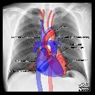
An anterosuperior mediastinal mass can be caused by neoplastic and non-neoplastic pathology. As their name suggests, they are confined to the anterior mediastinum, that portion of the mediastinum anterior to the pericardium and below the level of the clavicles.
The differential diagnosis for an anterior mediastinal mass includes:
- thymus
- thymoma: most common primary neoplasm of the anterosuperior mediastinum
- invasive thymoma
- thymic carcinoma
- thymolipoma/thymoliposarcoma
- thymic cyst
- thymic hyperplasia
- thymic carcinoid
- thyroid and parathyroid
- thyroid neoplasms
- thyroid goiter
- parathyroid neoplasms
- lymphoma
- germ cell tumors
- mediastinal teratoma
- mature: 75% of mediastinal germ cell tumors
- immature
- teratocarcinoma (malignant teratoma)
- mediastinal seminoma
- mediastinal embryonal cell carcinoma
- mediastinal yolk sac tumor
- mediastinal choriocarcinoma
- mediastinal mixed cell type germ cell tumor
- mediastinal teratoma
- thoracic aortic aneurysm
Mnemonic
- the 5 Ts
Siehe auch:
- Mediastinum
- Thymus
- Teratom
- Lymphom
- Perikard
- Chorionkarzinom
- Schilddrüse
- Keimzelltumor
- mediastinales Teratom
- Morbus Hodgkin
- thorakales Aortenaneurysma
- Normale Herzkonfiguration im Röntgen-Thorax
- anterosuperior mediastinal mass (mnemonic)
- vorderes Mediastinum
- Neoplasien der Schilddrüse
- Tumoren des Thymus
- mediastinales Seminom
- IgG4-assoziierte Erkrankung des Mediastinums
- hilum overlay sign
- chest radiograph in the exam setting
und weiter:
- Pancoast tumour
- Thymom
- Thymuskarzinom
- Morbus Castleman
- mediastinal lymphoma
- mediastinale Raumforderungen
- Thymuslipom
- enlargement of the cardiac silhouette
- retrosternal airspace
- invasives Thymom
- mediastinale Keimzelltumoren
- large-cell lymphoma of the mediastium
- obliteration of the retrosternal airspace
- oberes Mediastinum
- Raumforderungen oberes Mediastinum
- thymic epithelial tumours
- adult chest radiograph set-pieces
- Teratom des Thymus
- Metastasen im Thymus
- Karzinoid des Thymus
- mediastinal malignant germinoma
- lymphofollikuläre Thymushyperplasie
- hydatid cyst of the mediastinum
- Chondrosarkom des Sternums
- primäres Lymphom des Thymus

 Assoziationen und Differentialdiagnosen zu Tumoren des vorderen oberen Mediastinums:


