Trichilemmalzyste


Proliferating trichilemmal cysts, sometimes known as proliferating trichilemmal tumors, are dermal or subcutaneous tumors with squamoid cytologic features and trichilemmal-type keratinization, which usually arise in the scalp.
Terminology
A variety of names have been used for this pathology, including proliferating epidermoid cyst, pilar tumor of the scalp, proliferating epidermoid cyst, giant hair matrix tumor, hydatidiform keratinous cyst, trichochlamydocarcinoma, and invasive hair matrix tumor .
Epidemiology
Proliferating trichilemmal cysts have female predominance .
Associations
Some cases have a syndromic association:
Clinical presentation
Proliferating trichilemmal cysts present as lobulated masses within the scalp. They vary considerably in size from a few millimeters to large masses many centimeters in diameter. Occasionally clinical presentation will be with superimposed infection or malignant transformation, although both of these complications are uncommon .
Pathology
Although they are mostly benign, there are also local invasive and malignant types . The latter show extensive epithelial proliferation, variable cytologic atypia and mitotic activity.
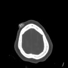
Radiographic features
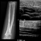
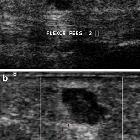
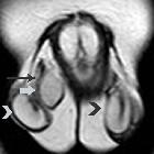
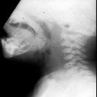
CT
Proliferating trichilemmal cysts usually are located within the scalp and appear as multiple complex subcutaneous solid or cystic masses. The cystic components contain high-density proteinaceous material which sometimes layers dependently . Ring-like patterns of mineralization are also encountered .
History and etymology
Proliferating trichilemmal cysts were first described by Edward Wilson Jones, an English dermatopathologist, in 1966 .
Differential diagnosis
In some situations, consider an epidermal inclusion cyst
See also
Siehe auch:
- intrakranielle Epidermoidzyste
- epidermale Inklusionszyste
- Ganglion (Überbein)
- Neurofibrom
- Epidermoid in der Kalotte
- Dermatofibrosarcoma protuberans
- Epidermoidzyste Finger
- testicular epidermoid cyst
- intraventricular epidermoid
- Atherom Galea
- epidermale Inklusionszyste der Mamma
- Fasciitis nodularis
- Epidermoidzyste vs Dermoidzyste
- kutane Metastasen
- Malherbe-Tumor
- atheroma
- reifes zystisches Teratom
und weiter:
- Mega Cisterna magna
- Arachnoidalzyste
- Rathke Zyste
- Cholesteatom
- ASP-Assoziation
- Pinealiszyste
- Ecchordosis physaliphora
- Gardner-Syndrom
- Tumor Kleinhirnbrückenwinkel
- Dermoidzyste
- Blake's-Pouch-Zyste
- Dandy-Walker continuum
- neuroglial cyst
- Keimzelltumor
- Akroosteolyse
- ependymal cyst
- erworbenes Cholesteatom
- Cholesteatom des äußeren Gehörgangs
- zystische Läsionen in der Hypophyse
- intraossäre Epidermoidzyste
- Steatocystoma multiplex
- hypothalamic lesions
- Dermoid Schädelkalotte
- testicular epidermoid
- congenital cholesteatoma
- white epidermoid
- periurethral cystic lesions
- Epidermoidzyste im Kleinhirnbrückenwinkel
- Epidermoid
- diffusionsgewichtete Bildgebung
- Galealipom
- Neoplasien der Cauda equina
- zystische Läsionen der Sellaregion
- Epidermoidzyste der Kalotte
- mostly / purely cystic pituitary region masses
- calcifying epithelioma of Malherbe
- mikrozystisches Meningeom
- diffusion MRI of an epidermoid tumor
- epidermoid cyst of the cauda equina
- Läsionen der Fingerspitze
- extratestikuläre intraskrotale Epidermoidzyste
- cystic extraaxial mass
- cutaneous epidermal cyst
- sutural epidermoid cyst
- Merkspruch Keimzelltumoren
- epidermale Inklusionszyste der Zunge
- Nasoalveoläre Zyste


 Assoziationen und Differentialdiagnosen zu epidermale Inklusionszyste: