Salter-Harris type I fracture

School ager
with ankle pain after a trampoline injury. AP (left) and oblique (right) radiographs of the ankle show a large amount of soft tissue swelling with its apex over the fibular physis. There is a transverse lucent line through the epiphysis of the fibula.The diagnosis was a non-displaced Salter-Harris fracture Type I of the distal fibula along with a fracture of the epiphysis of the fibula.

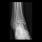
Teenager with
lateral malleolus pain after a motor vehicle accident. AP radiograph of the ankle shows a tremendous amount of swelling of the lateral malleolus with the apex of the swelling centered on the distal fibular physis. There is a small bony fragment near the physis as well thought to be from an avulsion injury.The diagnosis was a Salter-Harris Type I fracture of the distal fibula.

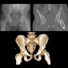
School ager
who tripped and fell, heard a pop, and then developed left hip pain. AP radiograph of the pelvis (upper left) shows the left femoral metaphysis to be displaced laterally from its epiphysis. This is better demonstrated on the coronal CT without contrast of the pelvis (upper right) and 3D CT of the pelvis (below)The diagnosis was slipped capital femoral epiphysis (Salter-Harris Type I fracture) of the left femur.
Salter-Harris type I fractures are relatively uncommon injuries that occur in children. Salter-Harris fractures are injuries where a fracture of the metaphysis or epiphysis extends through the physis. Not all fractures that extend to the growth plate are Salter-Harris fractures.
Radiographic features
Salter-Harris type I fractures describe a fracture that is completely contained within the physis. There is no associated bone fragment.
In reality, the majority of fractures that involve the physis have at least a small fragment of metaphysis associated with them and are therefore type II injuries.
Radiograph
- fracture through the physis
- no epiphyseal or metaphyseal fracture
- no fracture fragments
- angulation, displacement and rotation may occur
Related Radiopaedia articles
Fractures
- fracture
- terminology
- fracture location
- diaphyseal fracture
- metaphyseal fracture
- physeal fracture
- epiphyseal fracture
- fracture types
- avulsion fracture
- articular surface injuries
- complete fracture
- incomplete fracture
- infraction
- compound fracture
- pathological fracture
- stress fracture
- fracture displacement
- fracture translation
- fracture angulation
- fracture rotation
- fracture length
- distraction
- impaction
- shortening
- fracture location
- fracture healing
- skull fractures
- base of skull fractures
- skull vault fractures
- facial fractures
- fractures involving a single facial buttress
- alveolar process fractures
- frontal sinus fracture
- isolated zygomatic arch fractures
- mandibular fracture
- nasal bone fracture
- orbital blow-out fracture
- paranasal sinus fractures
- complex fractures
- dental fractures
- fractures involving a single facial buttress
- spinal fractures
- classification (AO Spine classification systems)
- cervical spine fracture classification systems
- AO classification of upper cervical injuries
- AO classification of subaxial injuries
- Anderson and D'Alonzo classification (odontoid fracture)
- Levine and Edwards classification (hangman fracture)
- Roy-Camille classification (odontoid process fracture )
- Allen and Ferguson classification (subaxial spine injuries)
- subaxial cervical spine injury classification (SLIC)
- thoracolumbar spinal fracture classification systems
- three column concept of spinal fractures (Denis classification)
- classification of sacral fractures
- cervical spine fracture classification systems
- spinal fractures by region
- spinal fracture types
- classification (AO Spine classification systems)
- rib fractures
- sternal fractures
- upper limb fractures
- classification
- Rockwood classification (acromioclavicular joint injury)
- AO classification (clavicle fracture)
- Neer classification (clavicle fracture)
- Neer classification (proximal humeral fracture)
- AO classification (proximal humeral fracture)
- AO/OTA classification of distal humeral fractures
- Milch classification (lateral humeral condyle fracture)
- Weiss classification (lateral humeral condyle fracture)
- Bado classification of Monteggia fracture-dislocations (radius-ulna)
- Mason classification (radial head fracture)
- Frykman classification (distal radial fracture)
- Mayo classification (scaphoid fracture)
- Hintermann classification (gamekeeper's thumb)
- Eaton classification (volar plate avulsion injury)
- Keifhaber-Stern classification (volar plate avulsion injury)
- upper limb fractures by region
- shoulder
- clavicular fracture
- scapular fracture
- acromion fracture
- coracoid process fracture
- glenoid fracture
- humeral head fracture
- proximal humeral fracture
- humeral neck fracture
- arm
- elbow
- forearm
- wrist
- carpal bones
- scaphoid fracture
- lunate fracture
- capitate fracture
- triquetral fracture
- pisiform fracture
- hamate fracture
- trapezoid fracture
- trapezium fracture
- hand
- shoulder
- classification
- lower limb fractures
- classification by region
- pelvis
- hip
- Pipkin classification (femoral head fracture)
- Garden classification (hip fracture)
- American Academy of Orthopedic Surgeons classification (periprosthetic hip fracture)
- Cooke and Newman classification (periprosthetic hip fracture)
- Johansson classification (periprosthetic hip fracture)
- Vancouver classification (periprosthetic hip fracture)
- femoral
- knee
- Schatzker classification (tibial plateau fracture)
- Meyers and McKeevers classification (anterior cruciate ligament avulsion fracture)
- tibia/fibula
- Watson-Jones classification (tibial tuberosity avulsion fracture)
- ankle
- foot
- Berndt and Harty classification (osteochondral lesions of the talus)
- Sanders CT classification (calcaneal fracture)
- Hawkins classification (talar neck fracture)
- Myerson classification (Lisfranc injury)
- Nunley-Vertullo classification (Lisfranc injury)
- pelvis and lower limb fractures by region
- pelvic fracture
- sacral fracture
- coccygeal fracture
- hip
- acetabular fracture
- femoral head fracture
- femoral neck fracture
- subcapital fracture
- transcervical fracture
- basicervical fracture
- trochanteric fracture
- pertrochanteric fracture
- intertrochanteric fracture
- subtrochanteric fracture
- thigh
- mid-shaft fracture
- bisphosphonate-related fracture
- knee
- avulsion fractures
- Segond fracture
- reverse Segond fracture
- anterior cruciate ligament avulsion fracture
- posterior cruciate ligament avulsion fracture
- arcuate complex avulsion fracture (arcuate sign)
- biceps femoris avulsion fracture
- iliotibial band avulsion fracture
- semimembranosus tendon avulsion fracture
- Stieda fracture (MCL avulsion fracture)
- patellar fracture
- tibial plateau fracture
- avulsion fractures
- leg
- tibial tuberosity avulsion fracture
- tibial shaft fracture
- fibular shaft fracture
- Maisonneuve fracture
- ankle
- foot
- tarsal bones
- Chopart fracture
- calcaneal fracture
- talar fracture
- navicular fracture
- medial cuneiform fracture
- intermediate cuneiform fracture
- lateral cuneiform fracture
- cuboid fracture
- metatarsal bones
- phalanges
- tarsal bones
- classification by region
- terminology
Siehe auch:

 Assoziationen und Differentialdiagnosen zu Salter-Harris Typ 1 Fraktur:


