Tumoren distales Femur

Teenager with
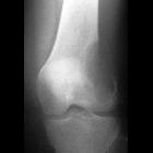
knee pain. AP radiograph of the knee shows growth plate fusion and a metaphyseal lesion that is lytic and expansile in appearance with a narrow zone of transition and no associated periosteal reaction.The diagnosis was giant cell tumor.

Enchondrom-typische
Läsion im distalen Femur als zufällige Nebenbefund Röntgenaufnahmen des Knies zur Planung einer Prothese bei medial betonter Gonarthrose.

A
biomechanical comparison between cement packing combined with extra fixation and three-dimensional printed strut-type prosthetic reconstruction for giant cell tumor of bone in distal femur. Postoperative T-SMART showed osseointegration: A a AP view of a 29 years olf male patient with GCTB. B Extended curettage, subchondral bone grafting and 3D-printed strut-type prosthetic reconstruction were performed. C T-SMART in postoperative day 1 showed interfacial gap between bone and implant (green box). D T-SMART taken at 2 years after surgery showed that excellent osseointegration

A
biomechanical comparison between cement packing combined with extra fixation and three-dimensional printed strut-type prosthetic reconstruction for giant cell tumor of bone in distal femur. Preoperative and postoperative X-ray evaluations: A a AP view of a 29 years old male patient with GCTB. B Extended curettage, cement packing, subchondral bone grafting and plate-screws fixation were performed. C A sclerotic rim occurred (green box), and an interfacial gap between bone and cement can be observed

Pitfalls in
the diagnosis of common benign bone tumours in children. Typical chondroblastoma of the distal femoral epiphysis: a Frontal X-ray of the right knee: well-defined round osteolytic lesion of the distal femoral epiphysis (arrow). b Sagittal fat-saturated proton density MRI: the lesion is well defined, mostly hyperintense, surrounded by bone oedema and an inflammatory reaction of the adjoining soft tissues. c CT of the right knee: the cortex is interrupted on the posterior border of the lesion (arrow), but no periosteal reaction is identified


Tumoren distales Femur
Knochentumoren Radiopaedia • CC-by-nc-sa 3.0 • de
There are a bewildering number of bone tumors with a wide variety of radiological appearances:
- bone-forming tumors
- cartilage-forming tumors
- fibrous bone lesions
- bone marrow tumors
- bony metastases
- other bone tumors or tumor-like lesions
- miscellaneous
- bone island / enostosis
- osteopoikilosis
- mastocytosis
- myeloid metaplasia-myelofibrosis
- pyknodysostosis
- radiation changes
- radiation-induced bone growth
- osteonecrosis
- radiation-induced bone tumors
See also
- soft tissue tumors
- WHO soft tissue tumor classification
- malignant fibrous histiocytoma
- liposarcoma
- synovial cell sarcoma
- fibromatoses
- rhabdomyosarcoma
- pigmented villonodular synovitis
- lipoma arborescens
Related Radiopaedia articles
Bone tumours
The differential diagnosis for bone tumors is dependent on the age of the patient, with a very different set of differentials for the pediatric patient.
- bone tumors
- bone-forming tumors
- cartilage-forming tumors
- fibrous bone lesions
- bone marrow tumors
- other bone tumors or tumor-like lesions
- adamantinoma
- aneurysmal bone cyst
- benign fibrous histiocytoma
- chordoma
- giant cell tumor of bone
- Gorham massive osteolysis
- hemangioendothelioma
- haemophilic pseudotumor
- intradiploic epidermoid cyst
- intraosseous lipoma
- musculoskeletal angiosarcoma
- musculoskeletal hemangiopericytoma
- primary intraosseous hemangioma
- post-traumatic cystic bone lesion
- simple bone cyst
- skeletal metastases
- morphology
- location
- impending fracture risk
- staging
- AJCC staging of musculoskeletal tumors
- Enneking surgical staging system
- approach
Siehe auch:
- Tumoren proximales Femur
- nicht ossifizierendes Fibrom distales Femur
- Enchondrom distales Femur
- eosinophiles Granulom Femur
- Knochentumoren des Femur
- parosteales Osteom
- Riesenzelltumor distales Femur
- Osteosarkom des distalen Femurs
- Chondroblastom distales Femur
- intraossäres Lipom des distalen Femurs
und weiter:

 Assoziationen und Differentialdiagnosen zu Tumoren distales Femur:


